Thomas Winkens, Anna Christl, Christian Kuehnel, Ferdinand Ndum, Philipp Seifert, Julia Greiser, Martin Freesmeyer
{"title":"鸵鸟蛋的卵内成像——计算机断层扫描对胚胎生理发育的评价","authors":"Thomas Winkens, Anna Christl, Christian Kuehnel, Ferdinand Ndum, Philipp Seifert, Julia Greiser, Martin Freesmeyer","doi":"10.1111/azo.12400","DOIUrl":null,"url":null,"abstract":"<p>In-ovo imaging using ostrich eggs has been described as a potential alternative to common animal testing. This approach is independent from small animal imaging devices as ostrich eggs provide good image quality on computed tomography (CT), MRI or PET scanners used in clinical routine examinations. However, questions regarding physiological development and systematic evaluation of image quality are open. This study aims at describing physiological development of ostrich embryos on serial CT scans. One hundred eggs (63 fertilized and 37 non-fertilized) were artificially incubated for 37 days. On developmental days (DD) 0, 10, 19, 22, 25, 28, 31, 34 and 37, CT scans were performed using a Siemens Biograph mCT40. Density of yolk, albumen and shell as well as volumes of air cell, egg content and egg shell were determined. In fertilized eggs, the size of different osseous structures was investigated. Detection of embryonal development was technically successful in 100%. Distinguishing of fertilized and non-fertilized eggs is achieved as early as DD 22. After that, continuous development is depicted and osseous structures become visible on DD 25. Ostrich eggs might open the door for preclinical imaging studies if small animal imaging devices are not available. This study contributes to the implementation of ostrich eggs as an alternative to common animal testing.</p>","PeriodicalId":50945,"journal":{"name":"Acta Zoologica","volume":"103 4","pages":"492-502"},"PeriodicalIF":1.1000,"publicationDate":"2021-08-03","publicationTypes":"Journal Article","fieldsOfStudy":null,"isOpenAccess":false,"openAccessPdf":"https://onlinelibrary.wiley.com/doi/epdf/10.1111/azo.12400","citationCount":"4","resultStr":"{\"title\":\"In-ovo imaging using ostrich eggs—Evaluation of physiological embryonal development on computed tomography\",\"authors\":\"Thomas Winkens, Anna Christl, Christian Kuehnel, Ferdinand Ndum, Philipp Seifert, Julia Greiser, Martin Freesmeyer\",\"doi\":\"10.1111/azo.12400\",\"DOIUrl\":null,\"url\":null,\"abstract\":\"<p>In-ovo imaging using ostrich eggs has been described as a potential alternative to common animal testing. This approach is independent from small animal imaging devices as ostrich eggs provide good image quality on computed tomography (CT), MRI or PET scanners used in clinical routine examinations. However, questions regarding physiological development and systematic evaluation of image quality are open. This study aims at describing physiological development of ostrich embryos on serial CT scans. One hundred eggs (63 fertilized and 37 non-fertilized) were artificially incubated for 37 days. On developmental days (DD) 0, 10, 19, 22, 25, 28, 31, 34 and 37, CT scans were performed using a Siemens Biograph mCT40. Density of yolk, albumen and shell as well as volumes of air cell, egg content and egg shell were determined. In fertilized eggs, the size of different osseous structures was investigated. Detection of embryonal development was technically successful in 100%. Distinguishing of fertilized and non-fertilized eggs is achieved as early as DD 22. After that, continuous development is depicted and osseous structures become visible on DD 25. Ostrich eggs might open the door for preclinical imaging studies if small animal imaging devices are not available. This study contributes to the implementation of ostrich eggs as an alternative to common animal testing.</p>\",\"PeriodicalId\":50945,\"journal\":{\"name\":\"Acta Zoologica\",\"volume\":\"103 4\",\"pages\":\"492-502\"},\"PeriodicalIF\":1.1000,\"publicationDate\":\"2021-08-03\",\"publicationTypes\":\"Journal Article\",\"fieldsOfStudy\":null,\"isOpenAccess\":false,\"openAccessPdf\":\"https://onlinelibrary.wiley.com/doi/epdf/10.1111/azo.12400\",\"citationCount\":\"4\",\"resultStr\":null,\"platform\":\"Semanticscholar\",\"paperid\":null,\"PeriodicalName\":\"Acta Zoologica\",\"FirstCategoryId\":\"99\",\"ListUrlMain\":\"https://onlinelibrary.wiley.com/doi/10.1111/azo.12400\",\"RegionNum\":4,\"RegionCategory\":\"生物学\",\"ArticlePicture\":[],\"TitleCN\":null,\"AbstractTextCN\":null,\"PMCID\":null,\"EPubDate\":\"\",\"PubModel\":\"\",\"JCR\":\"Q4\",\"JCRName\":\"ANATOMY & MORPHOLOGY\",\"Score\":null,\"Total\":0}","platform":"Semanticscholar","paperid":null,"PeriodicalName":"Acta Zoologica","FirstCategoryId":"99","ListUrlMain":"https://onlinelibrary.wiley.com/doi/10.1111/azo.12400","RegionNum":4,"RegionCategory":"生物学","ArticlePicture":[],"TitleCN":null,"AbstractTextCN":null,"PMCID":null,"EPubDate":"","PubModel":"","JCR":"Q4","JCRName":"ANATOMY & MORPHOLOGY","Score":null,"Total":0}
In-ovo imaging using ostrich eggs—Evaluation of physiological embryonal development on computed tomography
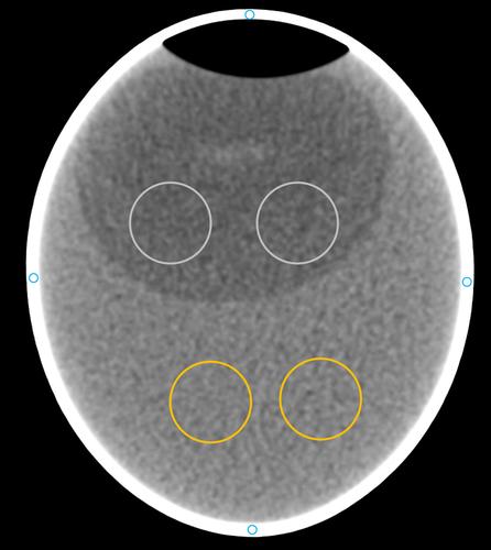
In-ovo imaging using ostrich eggs has been described as a potential alternative to common animal testing. This approach is independent from small animal imaging devices as ostrich eggs provide good image quality on computed tomography (CT), MRI or PET scanners used in clinical routine examinations. However, questions regarding physiological development and systematic evaluation of image quality are open. This study aims at describing physiological development of ostrich embryos on serial CT scans. One hundred eggs (63 fertilized and 37 non-fertilized) were artificially incubated for 37 days. On developmental days (DD) 0, 10, 19, 22, 25, 28, 31, 34 and 37, CT scans were performed using a Siemens Biograph mCT40. Density of yolk, albumen and shell as well as volumes of air cell, egg content and egg shell were determined. In fertilized eggs, the size of different osseous structures was investigated. Detection of embryonal development was technically successful in 100%. Distinguishing of fertilized and non-fertilized eggs is achieved as early as DD 22. After that, continuous development is depicted and osseous structures become visible on DD 25. Ostrich eggs might open the door for preclinical imaging studies if small animal imaging devices are not available. This study contributes to the implementation of ostrich eggs as an alternative to common animal testing.
期刊介绍:
Published regularly since 1920, Acta Zoologica has retained its position as one of the world''s leading journals in the field of animal organization, development, structure and function. Each issue publishes original research of interest to zoologists and physiologists worldwide, in the field of animal structure (from the cellular to the organismic level) and development with emphasis on functional, comparative and phylogenetic aspects. Occasional review articles are also published, as well as book reviews.


 分享
分享
 求助内容:
求助内容: 应助结果提醒方式:
 扫码关注我们
