{"title":"几何因子加权小波去噪在加多辛酸增强肝胆期MR成像中的应用。","authors":"Shota Kondo, Yuko Nakamura, Toru Higaki, Takashi Nishihara, Masahiro Takizawa, Toru Shirai, Motoshi Fujimori, Yoshitaka Bito, Keigo Narita, Takahiro Sueoka, Yukiko Honda, Chihiro Tani, Kazuo Awai","doi":"10.2463/mrms.mp.2022-0041","DOIUrl":null,"url":null,"abstract":"<p><strong>Purpose: </strong>The wavelet denoising with geometry factor weighting (g-denoising) method can reduce the image noise by adapting to spatially varying noise levels induced by parallel imaging. The aim of this study was to investigate the clinical applicability of g-denoising on hepatobiliary-phase (HBP) images with gadoxetic acid.</p><p><strong>Methods: </strong>We subjected 53 patients suspected of harboring hepatic neoplastic lesions to gadoxetic acid-enhanced HBP imaging with and without g-denoising (g<sup>+</sup>HBP and g<sup>-</sup>HBP). The matrix size was reduced for g<sup>+</sup>HBP images to avoid prolonging the scanning time. Two radiologists calculated the SNR, the portal vein-, and paraspinal muscle contrast-to-noise ratio (CNR) relative to the hepatic parenchyma (liver-to-portal vein- and liver-to-muscle CNR). Two other radiologists independently graded the sharpness of the liver edge, the visibility of intrahepatic vessels, the image noise, the homogeneity of liver parenchyma, and the overall image quality using a 5-point scale. Differences between g<sup>-</sup>HBP and g<sup>+</sup>HBP images were determined with the two-sided Wilcoxon signed-rank test.</p><p><strong>Results: </strong>The liver-to-portal- and liver-to-muscle CNR and the SNR were significantly higher on g<sup>+</sup>HBP- than g<sup>-</sup>HBP images (P < 0.01), as was the qualitative score for the image noise, homogeneity of liver parenchyma, and overall image quality (P < 0.01). Although there were no significant differences in the scores for the sharpness of the liver edge or the score assigned for the visibility of intrahepatic vessels (P = 0.05, 0.43), with g<sup>+</sup>HBP the score was lower in three patients for the sharpness of the liver edge and in six patients for the visibility of intrahepatic vessels.</p><p><strong>Conclusion: </strong>At gadoxetic acid-enhanced HBP imaging, g-denoising yielded a better image quality than conventional HBP imaging although the anatomic details may be degraded.</p>","PeriodicalId":18119,"journal":{"name":"Magnetic Resonance in Medical Sciences","volume":"22 2","pages":"241-252"},"PeriodicalIF":2.5000,"publicationDate":"2023-04-01","publicationTypes":"Journal Article","fieldsOfStudy":null,"isOpenAccess":false,"openAccessPdf":"https://ftp.ncbi.nlm.nih.gov/pub/pmc/oa_pdf/b0/52/mrms-22-241.PMC10086400.pdf","citationCount":"0","resultStr":"{\"title\":\"Utility of Wavelet Denoising with Geometry Factor Weighting for Gadoxetic Acid-enhanced Hepatobiliary-phase MR Imaging.\",\"authors\":\"Shota Kondo, Yuko Nakamura, Toru Higaki, Takashi Nishihara, Masahiro Takizawa, Toru Shirai, Motoshi Fujimori, Yoshitaka Bito, Keigo Narita, Takahiro Sueoka, Yukiko Honda, Chihiro Tani, Kazuo Awai\",\"doi\":\"10.2463/mrms.mp.2022-0041\",\"DOIUrl\":null,\"url\":null,\"abstract\":\"<p><strong>Purpose: </strong>The wavelet denoising with geometry factor weighting (g-denoising) method can reduce the image noise by adapting to spatially varying noise levels induced by parallel imaging. The aim of this study was to investigate the clinical applicability of g-denoising on hepatobiliary-phase (HBP) images with gadoxetic acid.</p><p><strong>Methods: </strong>We subjected 53 patients suspected of harboring hepatic neoplastic lesions to gadoxetic acid-enhanced HBP imaging with and without g-denoising (g<sup>+</sup>HBP and g<sup>-</sup>HBP). The matrix size was reduced for g<sup>+</sup>HBP images to avoid prolonging the scanning time. Two radiologists calculated the SNR, the portal vein-, and paraspinal muscle contrast-to-noise ratio (CNR) relative to the hepatic parenchyma (liver-to-portal vein- and liver-to-muscle CNR). Two other radiologists independently graded the sharpness of the liver edge, the visibility of intrahepatic vessels, the image noise, the homogeneity of liver parenchyma, and the overall image quality using a 5-point scale. Differences between g<sup>-</sup>HBP and g<sup>+</sup>HBP images were determined with the two-sided Wilcoxon signed-rank test.</p><p><strong>Results: </strong>The liver-to-portal- and liver-to-muscle CNR and the SNR were significantly higher on g<sup>+</sup>HBP- than g<sup>-</sup>HBP images (P < 0.01), as was the qualitative score for the image noise, homogeneity of liver parenchyma, and overall image quality (P < 0.01). Although there were no significant differences in the scores for the sharpness of the liver edge or the score assigned for the visibility of intrahepatic vessels (P = 0.05, 0.43), with g<sup>+</sup>HBP the score was lower in three patients for the sharpness of the liver edge and in six patients for the visibility of intrahepatic vessels.</p><p><strong>Conclusion: </strong>At gadoxetic acid-enhanced HBP imaging, g-denoising yielded a better image quality than conventional HBP imaging although the anatomic details may be degraded.</p>\",\"PeriodicalId\":18119,\"journal\":{\"name\":\"Magnetic Resonance in Medical Sciences\",\"volume\":\"22 2\",\"pages\":\"241-252\"},\"PeriodicalIF\":2.5000,\"publicationDate\":\"2023-04-01\",\"publicationTypes\":\"Journal Article\",\"fieldsOfStudy\":null,\"isOpenAccess\":false,\"openAccessPdf\":\"https://ftp.ncbi.nlm.nih.gov/pub/pmc/oa_pdf/b0/52/mrms-22-241.PMC10086400.pdf\",\"citationCount\":\"0\",\"resultStr\":null,\"platform\":\"Semanticscholar\",\"paperid\":null,\"PeriodicalName\":\"Magnetic Resonance in Medical Sciences\",\"FirstCategoryId\":\"3\",\"ListUrlMain\":\"https://doi.org/10.2463/mrms.mp.2022-0041\",\"RegionNum\":3,\"RegionCategory\":\"医学\",\"ArticlePicture\":[],\"TitleCN\":null,\"AbstractTextCN\":null,\"PMCID\":null,\"EPubDate\":\"\",\"PubModel\":\"\",\"JCR\":\"Q2\",\"JCRName\":\"RADIOLOGY, NUCLEAR MEDICINE & MEDICAL IMAGING\",\"Score\":null,\"Total\":0}","platform":"Semanticscholar","paperid":null,"PeriodicalName":"Magnetic Resonance in Medical Sciences","FirstCategoryId":"3","ListUrlMain":"https://doi.org/10.2463/mrms.mp.2022-0041","RegionNum":3,"RegionCategory":"医学","ArticlePicture":[],"TitleCN":null,"AbstractTextCN":null,"PMCID":null,"EPubDate":"","PubModel":"","JCR":"Q2","JCRName":"RADIOLOGY, NUCLEAR MEDICINE & MEDICAL IMAGING","Score":null,"Total":0}
Utility of Wavelet Denoising with Geometry Factor Weighting for Gadoxetic Acid-enhanced Hepatobiliary-phase MR Imaging.
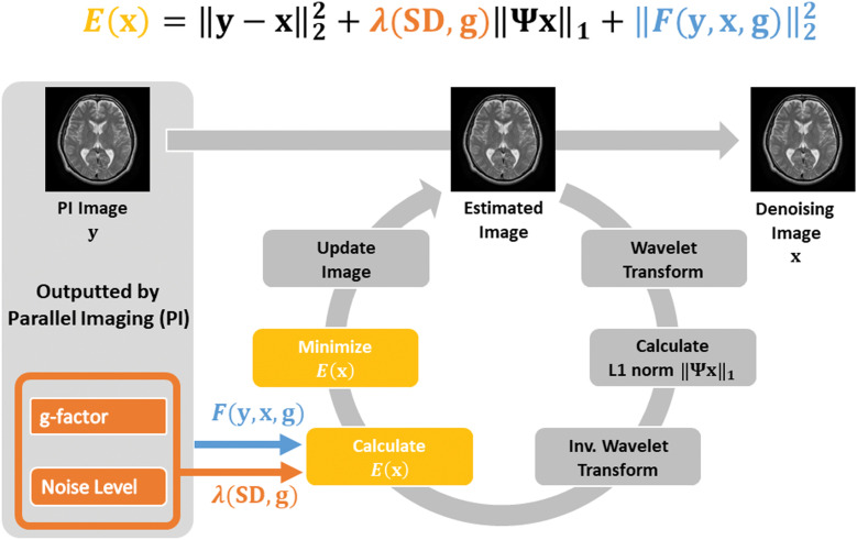
Purpose: The wavelet denoising with geometry factor weighting (g-denoising) method can reduce the image noise by adapting to spatially varying noise levels induced by parallel imaging. The aim of this study was to investigate the clinical applicability of g-denoising on hepatobiliary-phase (HBP) images with gadoxetic acid.
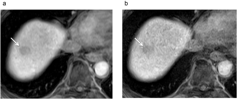
Methods: We subjected 53 patients suspected of harboring hepatic neoplastic lesions to gadoxetic acid-enhanced HBP imaging with and without g-denoising (g+HBP and g-HBP). The matrix size was reduced for g+HBP images to avoid prolonging the scanning time. Two radiologists calculated the SNR, the portal vein-, and paraspinal muscle contrast-to-noise ratio (CNR) relative to the hepatic parenchyma (liver-to-portal vein- and liver-to-muscle CNR). Two other radiologists independently graded the sharpness of the liver edge, the visibility of intrahepatic vessels, the image noise, the homogeneity of liver parenchyma, and the overall image quality using a 5-point scale. Differences between g-HBP and g+HBP images were determined with the two-sided Wilcoxon signed-rank test.
Results: The liver-to-portal- and liver-to-muscle CNR and the SNR were significantly higher on g+HBP- than g-HBP images (P < 0.01), as was the qualitative score for the image noise, homogeneity of liver parenchyma, and overall image quality (P < 0.01). Although there were no significant differences in the scores for the sharpness of the liver edge or the score assigned for the visibility of intrahepatic vessels (P = 0.05, 0.43), with g+HBP the score was lower in three patients for the sharpness of the liver edge and in six patients for the visibility of intrahepatic vessels.
Conclusion: At gadoxetic acid-enhanced HBP imaging, g-denoising yielded a better image quality than conventional HBP imaging although the anatomic details may be degraded.
期刊介绍:
Magnetic Resonance in Medical Sciences (MRMS or Magn
Reson Med Sci) is an international journal pursuing the
publication of original articles contributing to the progress
of magnetic resonance in the field of biomedical sciences
including technical developments and clinical applications.
MRMS is an official journal of the Japanese Society for
Magnetic Resonance in Medicine (JSMRM).


 分享
分享
 求助内容:
求助内容: 应助结果提醒方式:
 扫码关注我们
