Joseph DiPalma , Lorenzo Torresani , Saeed Hassanpour
{"title":"HistoPerm:一种基于排列的视图生成方法,用于改善组织病理特征表示学习","authors":"Joseph DiPalma , Lorenzo Torresani , Saeed Hassanpour","doi":"10.1016/j.jpi.2023.100320","DOIUrl":null,"url":null,"abstract":"<div><p>Deep learning has been effective for histology image analysis in digital pathology. However, many current deep learning approaches require large, strongly- or weakly labeled images and regions of interest, which can be time-consuming and resource-intensive to obtain. To address this challenge, we present HistoPerm, a view generation method for representation learning using joint embedding architectures that enhances representation learning for histology images. HistoPerm permutes augmented views of patches extracted from whole-slide histology images to improve classification performance. We evaluated the effectiveness of HistoPerm on 2 histology image datasets for Celiac disease and Renal Cell Carcinoma, using 3 widely used joint embedding architecture-based representation learning methods: BYOL, SimCLR, and VICReg. Our results show that HistoPerm consistently improves patch- and slide-level classification performance in terms of accuracy, F1-score, and AUC. Specifically, for patch-level classification accuracy on the Celiac disease dataset, HistoPerm boosts BYOL and VICReg by 8% and SimCLR by 3%. On the Renal Cell Carcinoma dataset, patch-level classification accuracy is increased by 2% for BYOL and VICReg, and by 1% for SimCLR. In addition, on the Celiac disease dataset, models with HistoPerm outperform the fully supervised baseline model by 6%, 5%, and 2% for BYOL, SimCLR, and VICReg, respectively. For the Renal Cell Carcinoma dataset, HistoPerm lowers the classification accuracy gap for the models up to 10% relative to the fully supervised baseline. These findings suggest that HistoPerm can be a valuable tool for improving representation learning of histopathology features when access to labeled data is limited and can lead to whole-slide classification results that are comparable to or superior to fully supervised methods.</p></div>","PeriodicalId":37769,"journal":{"name":"Journal of Pathology Informatics","volume":"14 ","pages":"Article 100320"},"PeriodicalIF":0.0000,"publicationDate":"2023-01-01","publicationTypes":"Journal Article","fieldsOfStudy":null,"isOpenAccess":false,"openAccessPdf":"https://www.ncbi.nlm.nih.gov/pmc/articles/PMC10339175/pdf/","citationCount":"2","resultStr":"{\"title\":\"HistoPerm: A permutation-based view generation approach for improving histopathologic feature representation learning\",\"authors\":\"Joseph DiPalma , Lorenzo Torresani , Saeed Hassanpour\",\"doi\":\"10.1016/j.jpi.2023.100320\",\"DOIUrl\":null,\"url\":null,\"abstract\":\"<div><p>Deep learning has been effective for histology image analysis in digital pathology. However, many current deep learning approaches require large, strongly- or weakly labeled images and regions of interest, which can be time-consuming and resource-intensive to obtain. To address this challenge, we present HistoPerm, a view generation method for representation learning using joint embedding architectures that enhances representation learning for histology images. HistoPerm permutes augmented views of patches extracted from whole-slide histology images to improve classification performance. We evaluated the effectiveness of HistoPerm on 2 histology image datasets for Celiac disease and Renal Cell Carcinoma, using 3 widely used joint embedding architecture-based representation learning methods: BYOL, SimCLR, and VICReg. Our results show that HistoPerm consistently improves patch- and slide-level classification performance in terms of accuracy, F1-score, and AUC. Specifically, for patch-level classification accuracy on the Celiac disease dataset, HistoPerm boosts BYOL and VICReg by 8% and SimCLR by 3%. On the Renal Cell Carcinoma dataset, patch-level classification accuracy is increased by 2% for BYOL and VICReg, and by 1% for SimCLR. In addition, on the Celiac disease dataset, models with HistoPerm outperform the fully supervised baseline model by 6%, 5%, and 2% for BYOL, SimCLR, and VICReg, respectively. For the Renal Cell Carcinoma dataset, HistoPerm lowers the classification accuracy gap for the models up to 10% relative to the fully supervised baseline. These findings suggest that HistoPerm can be a valuable tool for improving representation learning of histopathology features when access to labeled data is limited and can lead to whole-slide classification results that are comparable to or superior to fully supervised methods.</p></div>\",\"PeriodicalId\":37769,\"journal\":{\"name\":\"Journal of Pathology Informatics\",\"volume\":\"14 \",\"pages\":\"Article 100320\"},\"PeriodicalIF\":0.0000,\"publicationDate\":\"2023-01-01\",\"publicationTypes\":\"Journal Article\",\"fieldsOfStudy\":null,\"isOpenAccess\":false,\"openAccessPdf\":\"https://www.ncbi.nlm.nih.gov/pmc/articles/PMC10339175/pdf/\",\"citationCount\":\"2\",\"resultStr\":null,\"platform\":\"Semanticscholar\",\"paperid\":null,\"PeriodicalName\":\"Journal of Pathology Informatics\",\"FirstCategoryId\":\"1085\",\"ListUrlMain\":\"https://www.sciencedirect.com/science/article/pii/S2153353923001347\",\"RegionNum\":0,\"RegionCategory\":null,\"ArticlePicture\":[],\"TitleCN\":null,\"AbstractTextCN\":null,\"PMCID\":null,\"EPubDate\":\"2023/7/4 0:00:00\",\"PubModel\":\"Epub\",\"JCR\":\"Q2\",\"JCRName\":\"Medicine\",\"Score\":null,\"Total\":0}","platform":"Semanticscholar","paperid":null,"PeriodicalName":"Journal of Pathology Informatics","FirstCategoryId":"1085","ListUrlMain":"https://www.sciencedirect.com/science/article/pii/S2153353923001347","RegionNum":0,"RegionCategory":null,"ArticlePicture":[],"TitleCN":null,"AbstractTextCN":null,"PMCID":null,"EPubDate":"2023/7/4 0:00:00","PubModel":"Epub","JCR":"Q2","JCRName":"Medicine","Score":null,"Total":0}
HistoPerm: A permutation-based view generation approach for improving histopathologic feature representation learning
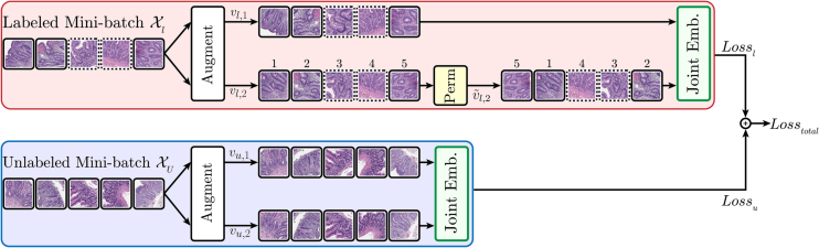
Deep learning has been effective for histology image analysis in digital pathology. However, many current deep learning approaches require large, strongly- or weakly labeled images and regions of interest, which can be time-consuming and resource-intensive to obtain. To address this challenge, we present HistoPerm, a view generation method for representation learning using joint embedding architectures that enhances representation learning for histology images. HistoPerm permutes augmented views of patches extracted from whole-slide histology images to improve classification performance. We evaluated the effectiveness of HistoPerm on 2 histology image datasets for Celiac disease and Renal Cell Carcinoma, using 3 widely used joint embedding architecture-based representation learning methods: BYOL, SimCLR, and VICReg. Our results show that HistoPerm consistently improves patch- and slide-level classification performance in terms of accuracy, F1-score, and AUC. Specifically, for patch-level classification accuracy on the Celiac disease dataset, HistoPerm boosts BYOL and VICReg by 8% and SimCLR by 3%. On the Renal Cell Carcinoma dataset, patch-level classification accuracy is increased by 2% for BYOL and VICReg, and by 1% for SimCLR. In addition, on the Celiac disease dataset, models with HistoPerm outperform the fully supervised baseline model by 6%, 5%, and 2% for BYOL, SimCLR, and VICReg, respectively. For the Renal Cell Carcinoma dataset, HistoPerm lowers the classification accuracy gap for the models up to 10% relative to the fully supervised baseline. These findings suggest that HistoPerm can be a valuable tool for improving representation learning of histopathology features when access to labeled data is limited and can lead to whole-slide classification results that are comparable to or superior to fully supervised methods.
期刊介绍:
The Journal of Pathology Informatics (JPI) is an open access peer-reviewed journal dedicated to the advancement of pathology informatics. This is the official journal of the Association for Pathology Informatics (API). The journal aims to publish broadly about pathology informatics and freely disseminate all articles worldwide. This journal is of interest to pathologists, informaticians, academics, researchers, health IT specialists, information officers, IT staff, vendors, and anyone with an interest in informatics. We encourage submissions from anyone with an interest in the field of pathology informatics. We publish all types of papers related to pathology informatics including original research articles, technical notes, reviews, viewpoints, commentaries, editorials, symposia, meeting abstracts, book reviews, and correspondence to the editors. All submissions are subject to rigorous peer review by the well-regarded editorial board and by expert referees in appropriate specialties.


 分享
分享
 求助内容:
求助内容: 应助结果提醒方式:
 扫码关注我们
