Konstantinos Natsis, George Tsakotos, George Triantafyllou, Łukasz Olewnik, Nicol Zielinska, Christos Koutserimpas, Trifon Totlis, Maria Piagkou
{"title":"手臂前后隔室的肌肉互连:一个可能具有临床意义的尸体病例系列。","authors":"Konstantinos Natsis, George Tsakotos, George Triantafyllou, Łukasz Olewnik, Nicol Zielinska, Christos Koutserimpas, Trifon Totlis, Maria Piagkou","doi":"10.1007/s00276-023-03209-5","DOIUrl":null,"url":null,"abstract":"<p><strong>Purpose: </strong>The report describes four cases of accessory bundles (ABs) or fibers connecting the muscles of the anterior with the posterior arm compartment. The ABs morphology (pure muscular or musculofascial or musculoaponeurotic) is described emphasizing their attachment points, characterized as muscles' interconnections.</p><p><strong>Materials and methods: </strong>Four formalin-embalmed donated male cadavers were dissected.</p><p><strong>Results: </strong>The muscles' interconnections were unilaterally identified. In the first case, the two ABs originated from the coracobrachialis muscle (CB), received fibers from the biceps brachii (BB), and were inserted into the triceps brachii (TB) medial head. The ABs created an arch over the brachial vessels and the median nerve (MN). In the second case, an accessory musculoaponeurotic structure was identified between CB and TB medial head and extended over the brachial vessels. In the third case, the myofascial ABs between the BB short head and the upper arm fascia, coursed anterior to the MN, the brachial artery, and the ulnar nerve, with direction to the TB medial head. In the fourth case, the three muscular ABs originating from the CB superficial and deep heads, in common with the BB short head, joined the upper arm fascia and the TB medial head and possibly entrapped the musculocutaneous nerve, the MN, and the brachial artery.</p><p><strong>Conclusion: </strong>ABs or musculoaponeurotic extensions may predispose to complications due to their potential compression on nerves and vessels. Clinicians should consider the possible existence of such bridging variants between muscles, in the differential diagnosis of a patient presenting with ischemia, edema, or MN palsy symptoms.</p>","PeriodicalId":49296,"journal":{"name":"Surgical and Radiologic Anatomy","volume":" ","pages":"1111-1116"},"PeriodicalIF":1.2000,"publicationDate":"2023-09-01","publicationTypes":"Journal Article","fieldsOfStudy":null,"isOpenAccess":false,"openAccessPdf":"https://www.ncbi.nlm.nih.gov/pmc/articles/PMC10514112/pdf/","citationCount":"0","resultStr":"{\"title\":\"Muscle interconnections in the anterior and posterior arm compartment: a cadaveric case series with possible clinical implications.\",\"authors\":\"Konstantinos Natsis, George Tsakotos, George Triantafyllou, Łukasz Olewnik, Nicol Zielinska, Christos Koutserimpas, Trifon Totlis, Maria Piagkou\",\"doi\":\"10.1007/s00276-023-03209-5\",\"DOIUrl\":null,\"url\":null,\"abstract\":\"<p><strong>Purpose: </strong>The report describes four cases of accessory bundles (ABs) or fibers connecting the muscles of the anterior with the posterior arm compartment. The ABs morphology (pure muscular or musculofascial or musculoaponeurotic) is described emphasizing their attachment points, characterized as muscles' interconnections.</p><p><strong>Materials and methods: </strong>Four formalin-embalmed donated male cadavers were dissected.</p><p><strong>Results: </strong>The muscles' interconnections were unilaterally identified. In the first case, the two ABs originated from the coracobrachialis muscle (CB), received fibers from the biceps brachii (BB), and were inserted into the triceps brachii (TB) medial head. The ABs created an arch over the brachial vessels and the median nerve (MN). In the second case, an accessory musculoaponeurotic structure was identified between CB and TB medial head and extended over the brachial vessels. In the third case, the myofascial ABs between the BB short head and the upper arm fascia, coursed anterior to the MN, the brachial artery, and the ulnar nerve, with direction to the TB medial head. In the fourth case, the three muscular ABs originating from the CB superficial and deep heads, in common with the BB short head, joined the upper arm fascia and the TB medial head and possibly entrapped the musculocutaneous nerve, the MN, and the brachial artery.</p><p><strong>Conclusion: </strong>ABs or musculoaponeurotic extensions may predispose to complications due to their potential compression on nerves and vessels. Clinicians should consider the possible existence of such bridging variants between muscles, in the differential diagnosis of a patient presenting with ischemia, edema, or MN palsy symptoms.</p>\",\"PeriodicalId\":49296,\"journal\":{\"name\":\"Surgical and Radiologic Anatomy\",\"volume\":\" \",\"pages\":\"1111-1116\"},\"PeriodicalIF\":1.2000,\"publicationDate\":\"2023-09-01\",\"publicationTypes\":\"Journal Article\",\"fieldsOfStudy\":null,\"isOpenAccess\":false,\"openAccessPdf\":\"https://www.ncbi.nlm.nih.gov/pmc/articles/PMC10514112/pdf/\",\"citationCount\":\"0\",\"resultStr\":null,\"platform\":\"Semanticscholar\",\"paperid\":null,\"PeriodicalName\":\"Surgical and Radiologic Anatomy\",\"FirstCategoryId\":\"3\",\"ListUrlMain\":\"https://doi.org/10.1007/s00276-023-03209-5\",\"RegionNum\":4,\"RegionCategory\":\"医学\",\"ArticlePicture\":[],\"TitleCN\":null,\"AbstractTextCN\":null,\"PMCID\":null,\"EPubDate\":\"2023/7/19 0:00:00\",\"PubModel\":\"Epub\",\"JCR\":\"Q3\",\"JCRName\":\"ANATOMY & MORPHOLOGY\",\"Score\":null,\"Total\":0}","platform":"Semanticscholar","paperid":null,"PeriodicalName":"Surgical and Radiologic Anatomy","FirstCategoryId":"3","ListUrlMain":"https://doi.org/10.1007/s00276-023-03209-5","RegionNum":4,"RegionCategory":"医学","ArticlePicture":[],"TitleCN":null,"AbstractTextCN":null,"PMCID":null,"EPubDate":"2023/7/19 0:00:00","PubModel":"Epub","JCR":"Q3","JCRName":"ANATOMY & MORPHOLOGY","Score":null,"Total":0}
Muscle interconnections in the anterior and posterior arm compartment: a cadaveric case series with possible clinical implications.
Purpose: The report describes four cases of accessory bundles (ABs) or fibers connecting the muscles of the anterior with the posterior arm compartment. The ABs morphology (pure muscular or musculofascial or musculoaponeurotic) is described emphasizing their attachment points, characterized as muscles' interconnections.
Materials and methods: Four formalin-embalmed donated male cadavers were dissected.
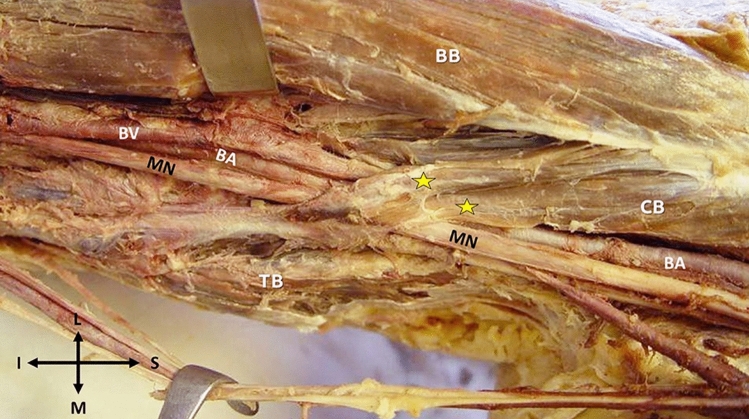
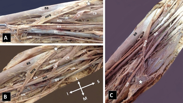
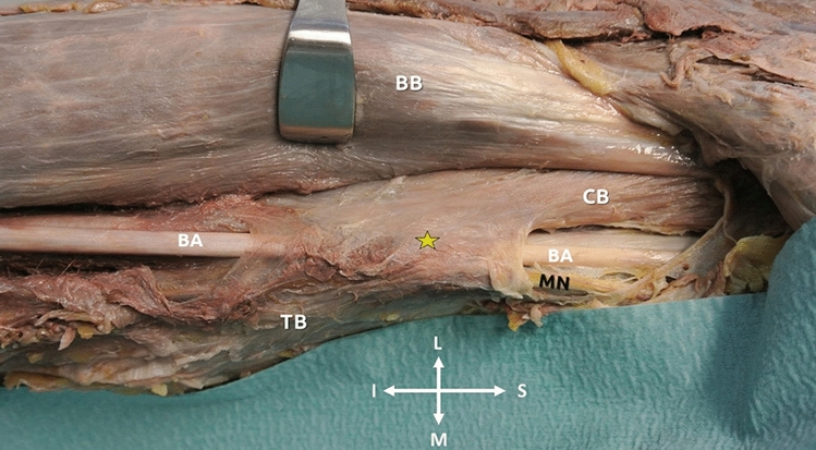
Results: The muscles' interconnections were unilaterally identified. In the first case, the two ABs originated from the coracobrachialis muscle (CB), received fibers from the biceps brachii (BB), and were inserted into the triceps brachii (TB) medial head. The ABs created an arch over the brachial vessels and the median nerve (MN). In the second case, an accessory musculoaponeurotic structure was identified between CB and TB medial head and extended over the brachial vessels. In the third case, the myofascial ABs between the BB short head and the upper arm fascia, coursed anterior to the MN, the brachial artery, and the ulnar nerve, with direction to the TB medial head. In the fourth case, the three muscular ABs originating from the CB superficial and deep heads, in common with the BB short head, joined the upper arm fascia and the TB medial head and possibly entrapped the musculocutaneous nerve, the MN, and the brachial artery.
Conclusion: ABs or musculoaponeurotic extensions may predispose to complications due to their potential compression on nerves and vessels. Clinicians should consider the possible existence of such bridging variants between muscles, in the differential diagnosis of a patient presenting with ischemia, edema, or MN palsy symptoms.
期刊介绍:
Anatomy is a morphological science which cannot fail to interest the clinician. The practical application of anatomical research to clinical problems necessitates special adaptation and selectivity in choosing from numerous international works. Although there is a tendency to believe that meaningful advances in anatomy are unlikely, constant revision is necessary. Surgical and Radiologic Anatomy, the first international journal of Clinical anatomy has been created in this spirit.
Its goal is to serve clinicians, regardless of speciality-physicians, surgeons, radiologists or other specialists-as an indispensable aid with which they can improve their knowledge of anatomy. Each issue includes: Original papers, review articles, articles on the anatomical bases of medical, surgical and radiological techniques, articles of normal radiologic anatomy, brief reviews of anatomical publications of clinical interest.
Particular attention is given to high quality illustrations, which are indispensable for a better understanding of anatomical problems.
Surgical and Radiologic Anatomy is a journal written by anatomists for clinicians with a special interest in anatomy.


 分享
分享
 求助内容:
求助内容: 应助结果提醒方式:
 扫码关注我们
