Barış Genç, Kerim Aslan, Ali Özçağlayan, Lütfi İncesu
{"title":"用高级弥散成像评估胶质母细胞瘤患者对侧正常白质的微结构异常。","authors":"Barış Genç, Kerim Aslan, Ali Özçağlayan, Lütfi İncesu","doi":"10.2463/mrms.mp.2023-0054","DOIUrl":null,"url":null,"abstract":"<p><strong>Purpose: </strong>Glioblastoma patients develop recurrence in the opposite hemisphere far from the primary tumor site even after complete resection. This is one of the main reasons for short disease survival. Our aim in this study is to detect microstructural changes in the contralateral hemisphere of glioblastoma patients using different diffusion models with the fully automated tract-based spatial statistics (TBSS) method.</p><p><strong>Methods: </strong>Fourteen right-sided and eleven left-sided glioblastoma patients without any treatment and eighteen age- and gender-matched controls were included in the study. Multi-shell diffusion weighted images were created with a 3T MRI device. After various preprocessing steps, images of fractional anisotropy (FA), mean diffusivity (MD), axial diffusivity (AD), radial diffusivity (RD), axial kurtosis (AK), mean kurtosis (MK), radial kurtosis (RK), intracellular volume fraction (ICVF), orientation dispersion index (ODI), and isotropic water fraction (ISO) were obtained. TBSS was used to compare diffusion tensor imaging, diffusion kurtosis imaging, and neurite orientation dispersion and density imaging parameters of right- and left-sided glioblastoma patients with the control group for the contralateral hemisphere.</p><p><strong>Results: </strong>Both right-sided and left-sided glioblastoma patients have shown an increase in MD and ODI in the contralateral hemisphere. While right-sided glioblastoma patients showed an increase in RD, AD, and ISO in a more limited area in the contralateral hemisphere, left-sided glioblastoma patients showed an increase in MK and AK. FA, ICVF, and RK did not show any difference in both groups.</p><p><strong>Conclusion: </strong>There are microstructural changes in the contralateral hemisphere in glioblastoma patients, and these changes differ between right-sided and left-sided glioblastoma patients.</p>","PeriodicalId":18119,"journal":{"name":"Magnetic Resonance in Medical Sciences","volume":" ","pages":"479-486"},"PeriodicalIF":3.2000,"publicationDate":"2024-10-01","publicationTypes":"Journal Article","fieldsOfStudy":null,"isOpenAccess":false,"openAccessPdf":"https://www.ncbi.nlm.nih.gov/pmc/articles/PMC11447469/pdf/","citationCount":"0","resultStr":"{\"title\":\"Microstructural Abnormalities in the Contralateral Normal-appearing White Matter of Glioblastoma Patients Evaluated with Advanced Diffusion Imaging.\",\"authors\":\"Barış Genç, Kerim Aslan, Ali Özçağlayan, Lütfi İncesu\",\"doi\":\"10.2463/mrms.mp.2023-0054\",\"DOIUrl\":null,\"url\":null,\"abstract\":\"<p><strong>Purpose: </strong>Glioblastoma patients develop recurrence in the opposite hemisphere far from the primary tumor site even after complete resection. This is one of the main reasons for short disease survival. Our aim in this study is to detect microstructural changes in the contralateral hemisphere of glioblastoma patients using different diffusion models with the fully automated tract-based spatial statistics (TBSS) method.</p><p><strong>Methods: </strong>Fourteen right-sided and eleven left-sided glioblastoma patients without any treatment and eighteen age- and gender-matched controls were included in the study. Multi-shell diffusion weighted images were created with a 3T MRI device. After various preprocessing steps, images of fractional anisotropy (FA), mean diffusivity (MD), axial diffusivity (AD), radial diffusivity (RD), axial kurtosis (AK), mean kurtosis (MK), radial kurtosis (RK), intracellular volume fraction (ICVF), orientation dispersion index (ODI), and isotropic water fraction (ISO) were obtained. TBSS was used to compare diffusion tensor imaging, diffusion kurtosis imaging, and neurite orientation dispersion and density imaging parameters of right- and left-sided glioblastoma patients with the control group for the contralateral hemisphere.</p><p><strong>Results: </strong>Both right-sided and left-sided glioblastoma patients have shown an increase in MD and ODI in the contralateral hemisphere. While right-sided glioblastoma patients showed an increase in RD, AD, and ISO in a more limited area in the contralateral hemisphere, left-sided glioblastoma patients showed an increase in MK and AK. FA, ICVF, and RK did not show any difference in both groups.</p><p><strong>Conclusion: </strong>There are microstructural changes in the contralateral hemisphere in glioblastoma patients, and these changes differ between right-sided and left-sided glioblastoma patients.</p>\",\"PeriodicalId\":18119,\"journal\":{\"name\":\"Magnetic Resonance in Medical Sciences\",\"volume\":\" \",\"pages\":\"479-486\"},\"PeriodicalIF\":3.2000,\"publicationDate\":\"2024-10-01\",\"publicationTypes\":\"Journal Article\",\"fieldsOfStudy\":null,\"isOpenAccess\":false,\"openAccessPdf\":\"https://www.ncbi.nlm.nih.gov/pmc/articles/PMC11447469/pdf/\",\"citationCount\":\"0\",\"resultStr\":null,\"platform\":\"Semanticscholar\",\"paperid\":null,\"PeriodicalName\":\"Magnetic Resonance in Medical Sciences\",\"FirstCategoryId\":\"3\",\"ListUrlMain\":\"https://doi.org/10.2463/mrms.mp.2023-0054\",\"RegionNum\":3,\"RegionCategory\":\"医学\",\"ArticlePicture\":[],\"TitleCN\":null,\"AbstractTextCN\":null,\"PMCID\":null,\"EPubDate\":\"2023/8/1 0:00:00\",\"PubModel\":\"Epub\",\"JCR\":\"Q2\",\"JCRName\":\"RADIOLOGY, NUCLEAR MEDICINE & MEDICAL IMAGING\",\"Score\":null,\"Total\":0}","platform":"Semanticscholar","paperid":null,"PeriodicalName":"Magnetic Resonance in Medical Sciences","FirstCategoryId":"3","ListUrlMain":"https://doi.org/10.2463/mrms.mp.2023-0054","RegionNum":3,"RegionCategory":"医学","ArticlePicture":[],"TitleCN":null,"AbstractTextCN":null,"PMCID":null,"EPubDate":"2023/8/1 0:00:00","PubModel":"Epub","JCR":"Q2","JCRName":"RADIOLOGY, NUCLEAR MEDICINE & MEDICAL IMAGING","Score":null,"Total":0}
引用次数: 0
摘要
目的:胶质母细胞瘤患者即使在完全切除肿瘤后,仍会在远离原发肿瘤部位的对侧半球复发。这是导致患者生存期短的主要原因之一。本研究的目的是利用不同的弥散模型和全自动基于束的空间统计(TBSS)方法检测胶质母细胞瘤患者对侧半球的微结构变化:研究对象包括 14 名未接受任何治疗的右侧和 11 名左侧胶质母细胞瘤患者,以及 18 名年龄和性别匹配的对照组患者。使用 3T 磁共振成像设备生成多壳弥散加权图像。经过各种预处理步骤后,获得了分数各向异性(FA)、平均扩散率(MD)、轴向扩散率(AD)、径向扩散率(RD)、轴向峰度(AK)、平均峰度(MK)、径向峰度(RK)、细胞内体积分数(ICVF)、方向弥散指数(ODI)和各向同性水分数(ISO)的图像。使用 TBSS 比较右侧和左侧胶质母细胞瘤患者与对侧半球对照组的弥散张量成像、弥散峰度成像、神经元取向弥散和密度成像参数:结果:右侧和左侧胶质母细胞瘤患者对侧半球的MD和ODI均有所增加。右侧胶质母细胞瘤患者的 RD、AD 和 ISO 在对侧半球更有限的区域内增加,而左侧胶质母细胞瘤患者的 MK 和 AK 增加。两组患者的FA、ICVF和RK没有任何差异:结论:胶质母细胞瘤患者的对侧大脑半球存在微结构变化,右侧和左侧胶质母细胞瘤患者的这些变化有所不同。
Microstructural Abnormalities in the Contralateral Normal-appearing White Matter of Glioblastoma Patients Evaluated with Advanced Diffusion Imaging.
Purpose: Glioblastoma patients develop recurrence in the opposite hemisphere far from the primary tumor site even after complete resection. This is one of the main reasons for short disease survival. Our aim in this study is to detect microstructural changes in the contralateral hemisphere of glioblastoma patients using different diffusion models with the fully automated tract-based spatial statistics (TBSS) method.
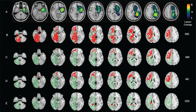
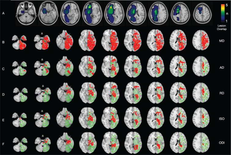
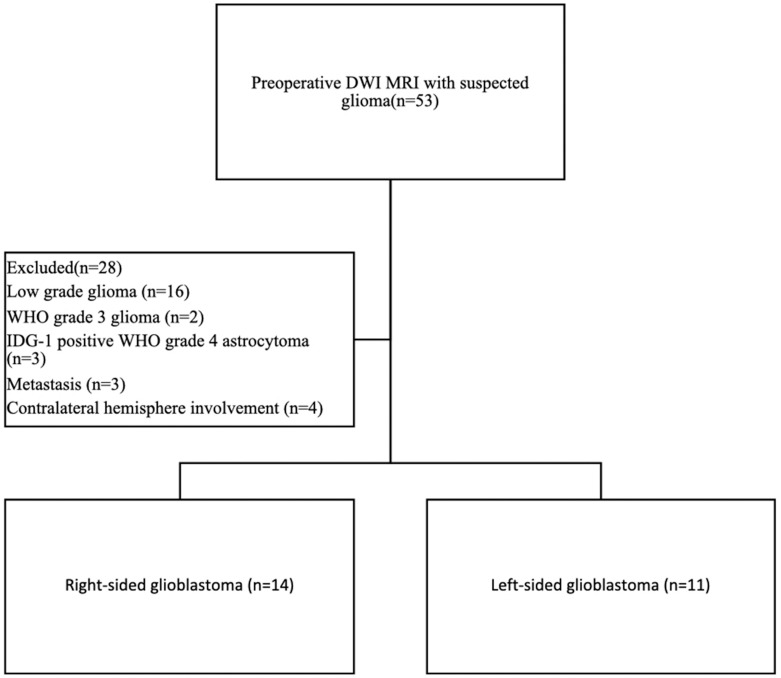
Methods: Fourteen right-sided and eleven left-sided glioblastoma patients without any treatment and eighteen age- and gender-matched controls were included in the study. Multi-shell diffusion weighted images were created with a 3T MRI device. After various preprocessing steps, images of fractional anisotropy (FA), mean diffusivity (MD), axial diffusivity (AD), radial diffusivity (RD), axial kurtosis (AK), mean kurtosis (MK), radial kurtosis (RK), intracellular volume fraction (ICVF), orientation dispersion index (ODI), and isotropic water fraction (ISO) were obtained. TBSS was used to compare diffusion tensor imaging, diffusion kurtosis imaging, and neurite orientation dispersion and density imaging parameters of right- and left-sided glioblastoma patients with the control group for the contralateral hemisphere.
Results: Both right-sided and left-sided glioblastoma patients have shown an increase in MD and ODI in the contralateral hemisphere. While right-sided glioblastoma patients showed an increase in RD, AD, and ISO in a more limited area in the contralateral hemisphere, left-sided glioblastoma patients showed an increase in MK and AK. FA, ICVF, and RK did not show any difference in both groups.
Conclusion: There are microstructural changes in the contralateral hemisphere in glioblastoma patients, and these changes differ between right-sided and left-sided glioblastoma patients.
期刊介绍:
Magnetic Resonance in Medical Sciences (MRMS or Magn
Reson Med Sci) is an international journal pursuing the
publication of original articles contributing to the progress
of magnetic resonance in the field of biomedical sciences
including technical developments and clinical applications.
MRMS is an official journal of the Japanese Society for
Magnetic Resonance in Medicine (JSMRM).


 分享
分享
 求助内容:
求助内容: 应助结果提醒方式:
 扫码关注我们
