To compare the application of different treatments in the diagnosis of melanoma with severe pigment interference, to solve the problem of pigment interference with immunohistochemical interpretation.
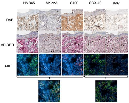
The pigment-rich melanomas were first depigmented with potassium permanganate using a concentration gradient (0.1%, 0.5%, 1%) and a time gradient (1, 5, 10, 15, 30 min, 6 h), and the optimal concentration and time were found. Then, 12 cases of pigment-rich melanoma tissues were collected, and the tissues were stained with diaminobenzidine (DAB), alkaline phosphatase-fast red (AP red), multiplex immunofluorescence (MIF), and 3-amino-9-ethylcarbazole (AEC), and ferrous sulfate, comparing different methods, positive expression of HMB45, MelanA, S100, SOX10, ki67.
First, the concentration of 0.5% potassium permanganate after 15 min treatment of the pigment significantly faded, and the intensity of antibody positivity was better than other concentrations and time. Second, after depigmentation treatment, the antibody positivity rate was 41.7%–66.7% for DAB, 66.7%–91.7% for AP red, 83.3%–100% for multiplex immunofluorescence, 25%–33.3% for AEC, and 33.3% for ferrous sulfate.
AP red staining and mIF are more suitable for the diagnosis of melanoma with severe pigment interference, and AP red staining is more economical and practical.


 分享
分享
 求助内容:
求助内容: 应助结果提醒方式:
 扫码关注我们
