Yue Li, Yunpeng Wang, Yuanyuan Ding, Xi Fan, Liansong Ye, Qingqing Pan, Bowen Zhang, Peng Li, Kui Luo, Bing Hu, Bin He, Yuji Pu
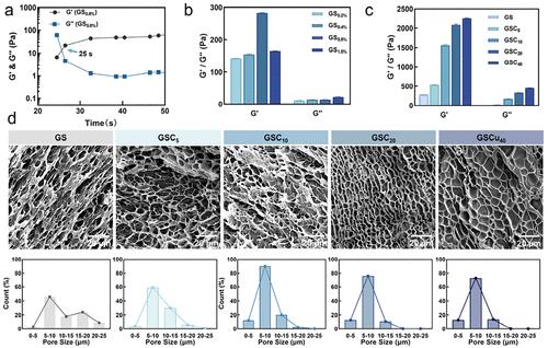
{"title":"Correction to “A Double Network Composite Hydrogel with Self-Regulating Cu2+/Luteolin Release and Mechanical Modulation for Enhanced Wound Healing”","authors":"Yue Li, Yunpeng Wang, Yuanyuan Ding, Xi Fan, Liansong Ye, Qingqing Pan, Bowen Zhang, Peng Li, Kui Luo, Bing Hu, Bin He, Yuji Pu","doi":"10.1021/acsnano.4c14461","DOIUrl":null,"url":null,"abstract":"In Figure 2d, the SEM of the GSC<sub>10</sub> was incorrectly used. We have replaced it with the correct image and recalculated the pore size distributions. Figure 2. Characterization of blank GS and GSC hydrogels. (a–c) Rheological properties of (a) GS0.8% with time after UV irradiation; (b) GS hydrogels prepared with different SA concentrations at ω = 10 rad/s; GSC hydrogels prepared with various Cu<sup>2+</sup> concentrations at ω = 10 rad/s. (d) SEM images and pore size distributions of GSC hydrogels. In Figure 6a, there was a typo in the group label. The group name “GC/PBE@Lut” should have been “GS/PBE@Lut.” We have corrected the figure accordingly. Figure 6. In vitro wound healing and angiogenesis. (a) Scratch assay assessing the proliferation and migration abilities of HUVEC after coculturing with different materials. (b) Quantification of wound closure ratios in the scratch assay. (c) Images of tube formation by HUVECs after coincubation with hydrogel extracts for 6 h. Quantifications of nodes (d) and tube length (e) in the tube formation assay. (f) RT-qPCR analysis of VEGF expression in HUVECs. *<i>p</i> < 0.05, **<i>p</i> < 0.01, and ***<i>p</i> < 0.001. Similarly, in the results and discussion sections 2.4, 2.5, and 2.7, GC/PBE@Lut should be changed into GS/PBE@Lut. The revised text is as follows: In Section 2.4: “The antibacterial efficacies of GSC, GS/PBE@Lut, and GSC/PBE@Lut against <i>Escherichia coli</i> and <i>Staphylococcus aureus</i> were investigated using a coculture method (Figure 4d).” In section 2.5: “To examine the effect of GS/PBE@Lut and GSC/PBE@Lut on cell proliferation and migration in chronic wound environments, a scratch assay was conducted under conditions of pH 7.4 and high oxidative stress induced by exogenous 100 μM H<sub>2</sub>O<sub>2</sub>.” “The results indicated a substantial enhancement in cell proliferation and migration for GSC, GS/PBE@Lut, and GSC/PBE@Lut compared with the H<sub>2</sub>O<sub>2</sub> group (Figure 6a).” “Promoting vascular regeneration is paramount to tissue regeneration. Following a 6 h coculture of HUVECs with extracts from the control group, GSC, GS/PBE@Lut, and GSC/PBE@Lut, the number of nodes and tube lengths were measured (Figure 6c). ” In Section 2.7: “The fluorescence intensities of GS/PBE@Lut and GSC/PBE@Lut were notably lower than those of the control group (Figure 9b,d).” “Quantitative analysis of CD206+/CD68+ cell percentages indicated 2.95- and 0.94-fold increases in M2 polarization for GSC/PBE@Lut and GS/PBE@Lut treatments, respectively (Figure 9e).” “Quantitative analysis of iNOS+/CD68+ cell percentages revealed no M1 macrophages in the GSC/PBE@Lut and GS/PBE@Lut groups, while the control and GSC groups exhibited M1 macrophages (Figure 9f).” In Figure 8, the skin section image of the GS group on day 15 was incorrectly used. We have replaced it with the correct image. Figure 8. (a) H&E staining of the wound tissue of days 5, 10, and 15; scale bar 100 μm. (b) Masson staining of the wound tissue; scale bar 1000 μm. Quantifications of collagen content (c) and epidermal thickness (d). *<i>p</i> < 0.05 and **<i>p</i> < 0.01. These are unintentional errors and do not affect the scientific conclusions corresponding to the main text of the article. All coauthors have approved this correction. We express our profound apologies for the inconvenience caused by our negligence to the editor and readers. This article has not yet been cited by other publications.","PeriodicalId":21,"journal":{"name":"ACS Nano","volume":"21 1","pages":""},"PeriodicalIF":16.0000,"publicationDate":"2024-12-03","publicationTypes":"Journal Article","fieldsOfStudy":null,"isOpenAccess":false,"openAccessPdf":"","citationCount":"0","resultStr":null,"platform":"Semanticscholar","paperid":null,"PeriodicalName":"ACS Nano","FirstCategoryId":"88","ListUrlMain":"https://doi.org/10.1021/acsnano.4c14461","RegionNum":1,"RegionCategory":"材料科学","ArticlePicture":[],"TitleCN":null,"AbstractTextCN":null,"PMCID":null,"EPubDate":"","PubModel":"","JCR":"Q1","JCRName":"CHEMISTRY, MULTIDISCIPLINARY","Score":null,"Total":0}
引用次数: 0
Abstract
In Figure 2d, the SEM of the GSC10 was incorrectly used. We have replaced it with the correct image and recalculated the pore size distributions. Figure 2. Characterization of blank GS and GSC hydrogels. (a–c) Rheological properties of (a) GS0.8% with time after UV irradiation; (b) GS hydrogels prepared with different SA concentrations at ω = 10 rad/s; GSC hydrogels prepared with various Cu2+ concentrations at ω = 10 rad/s. (d) SEM images and pore size distributions of GSC hydrogels. In Figure 6a, there was a typo in the group label. The group name “GC/PBE@Lut” should have been “GS/PBE@Lut.” We have corrected the figure accordingly. Figure 6. In vitro wound healing and angiogenesis. (a) Scratch assay assessing the proliferation and migration abilities of HUVEC after coculturing with different materials. (b) Quantification of wound closure ratios in the scratch assay. (c) Images of tube formation by HUVECs after coincubation with hydrogel extracts for 6 h. Quantifications of nodes (d) and tube length (e) in the tube formation assay. (f) RT-qPCR analysis of VEGF expression in HUVECs. *p < 0.05, **p < 0.01, and ***p < 0.001. Similarly, in the results and discussion sections 2.4, 2.5, and 2.7, GC/PBE@Lut should be changed into GS/PBE@Lut. The revised text is as follows: In Section 2.4: “The antibacterial efficacies of GSC, GS/PBE@Lut, and GSC/PBE@Lut against Escherichia coli and Staphylococcus aureus were investigated using a coculture method (Figure 4d).” In section 2.5: “To examine the effect of GS/PBE@Lut and GSC/PBE@Lut on cell proliferation and migration in chronic wound environments, a scratch assay was conducted under conditions of pH 7.4 and high oxidative stress induced by exogenous 100 μM H2O2.” “The results indicated a substantial enhancement in cell proliferation and migration for GSC, GS/PBE@Lut, and GSC/PBE@Lut compared with the H2O2 group (Figure 6a).” “Promoting vascular regeneration is paramount to tissue regeneration. Following a 6 h coculture of HUVECs with extracts from the control group, GSC, GS/PBE@Lut, and GSC/PBE@Lut, the number of nodes and tube lengths were measured (Figure 6c). ” In Section 2.7: “The fluorescence intensities of GS/PBE@Lut and GSC/PBE@Lut were notably lower than those of the control group (Figure 9b,d).” “Quantitative analysis of CD206+/CD68+ cell percentages indicated 2.95- and 0.94-fold increases in M2 polarization for GSC/PBE@Lut and GS/PBE@Lut treatments, respectively (Figure 9e).” “Quantitative analysis of iNOS+/CD68+ cell percentages revealed no M1 macrophages in the GSC/PBE@Lut and GS/PBE@Lut groups, while the control and GSC groups exhibited M1 macrophages (Figure 9f).” In Figure 8, the skin section image of the GS group on day 15 was incorrectly used. We have replaced it with the correct image. Figure 8. (a) H&E staining of the wound tissue of days 5, 10, and 15; scale bar 100 μm. (b) Masson staining of the wound tissue; scale bar 1000 μm. Quantifications of collagen content (c) and epidermal thickness (d). *p < 0.05 and **p < 0.01. These are unintentional errors and do not affect the scientific conclusions corresponding to the main text of the article. All coauthors have approved this correction. We express our profound apologies for the inconvenience caused by our negligence to the editor and readers. This article has not yet been cited by other publications.
期刊介绍:
ACS Nano, published monthly, serves as an international forum for comprehensive articles on nanoscience and nanotechnology research at the intersections of chemistry, biology, materials science, physics, and engineering. The journal fosters communication among scientists in these communities, facilitating collaboration, new research opportunities, and advancements through discoveries. ACS Nano covers synthesis, assembly, characterization, theory, and simulation of nanostructures, nanobiotechnology, nanofabrication, methods and tools for nanoscience and nanotechnology, and self- and directed-assembly. Alongside original research articles, it offers thorough reviews, perspectives on cutting-edge research, and discussions envisioning the future of nanoscience and nanotechnology.


 分享
分享
 求助内容:
求助内容: 应助结果提醒方式:
 扫码关注我们
