Lei Li, Xiaotong He, Yang Zhang, Dashan Qi, Meixing Li, Hui Zhang, Qingming Shen, Quli Fan
{"title":"Near-Infrared Light-Activated DNA Nanodevice for Spatiotemporal In Vivo Fluorescence Imaging of Messenger RNA","authors":"Lei Li, Xiaotong He, Yang Zhang, Dashan Qi, Meixing Li, Hui Zhang, Qingming Shen, Quli Fan","doi":"10.1021/acs.analchem.4c05292","DOIUrl":null,"url":null,"abstract":"Real-time visualization of messenger RNA (mRNA) is essential for tumor classification, grading, and staging. However, the low signal-to-background ratios and nonspatiotemporal specific signal amplification restricted the in vivo imaging of mRNA. In this study, a near-infrared (NIR) light-activated DNA nanodevice (DND) was developed for spatiotemporal in vivo fluorescence imaging of mRNA. The DND was fabricated by encapsulating indocyanine green (ICG) and DNA fluorescent probes within thermosensitive liposomes and subsequently functionalizing the liposomes with aptamers. The ICG offers the “always-on” fluorescence signal, offering a feasible strategy for monitoring DND distribution. The fluorescence signal of DNA probes remains inactive (“off” state) during the delivery process. Upon targeted delivery of the DNDs to tumor cells via aptamer recognition, the thermosensitive liposomes could be dissociated by the photothermal effect induced by ICG under near-infrared irradiation, thereby facilitating the release of DNA probes. The DNA probes were activated (“turn on”) by tumor-specific thymidine kinase 1 (TK1) mRNA through toehold-mediated strand displacement cascades, enabling the signal-amplified fluorescence imaging of mRNA. This study reveals the distinctive light-activated merit and remarkable fluorescence imaging of DNDs, highlighting their great potential to promote progress in spatiotemporal resolution imaging of other disease-relevant RNAs in vivo.","PeriodicalId":27,"journal":{"name":"Analytical Chemistry","volume":"27 1","pages":""},"PeriodicalIF":6.7000,"publicationDate":"2024-12-21","publicationTypes":"Journal Article","fieldsOfStudy":null,"isOpenAccess":false,"openAccessPdf":"","citationCount":"0","resultStr":null,"platform":"Semanticscholar","paperid":null,"PeriodicalName":"Analytical Chemistry","FirstCategoryId":"92","ListUrlMain":"https://doi.org/10.1021/acs.analchem.4c05292","RegionNum":1,"RegionCategory":"化学","ArticlePicture":[],"TitleCN":null,"AbstractTextCN":null,"PMCID":null,"EPubDate":"","PubModel":"","JCR":"Q1","JCRName":"CHEMISTRY, ANALYTICAL","Score":null,"Total":0}
引用次数: 0
Abstract
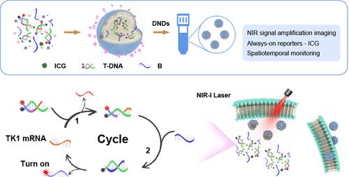
Real-time visualization of messenger RNA (mRNA) is essential for tumor classification, grading, and staging. However, the low signal-to-background ratios and nonspatiotemporal specific signal amplification restricted the in vivo imaging of mRNA. In this study, a near-infrared (NIR) light-activated DNA nanodevice (DND) was developed for spatiotemporal in vivo fluorescence imaging of mRNA. The DND was fabricated by encapsulating indocyanine green (ICG) and DNA fluorescent probes within thermosensitive liposomes and subsequently functionalizing the liposomes with aptamers. The ICG offers the “always-on” fluorescence signal, offering a feasible strategy for monitoring DND distribution. The fluorescence signal of DNA probes remains inactive (“off” state) during the delivery process. Upon targeted delivery of the DNDs to tumor cells via aptamer recognition, the thermosensitive liposomes could be dissociated by the photothermal effect induced by ICG under near-infrared irradiation, thereby facilitating the release of DNA probes. The DNA probes were activated (“turn on”) by tumor-specific thymidine kinase 1 (TK1) mRNA through toehold-mediated strand displacement cascades, enabling the signal-amplified fluorescence imaging of mRNA. This study reveals the distinctive light-activated merit and remarkable fluorescence imaging of DNDs, highlighting their great potential to promote progress in spatiotemporal resolution imaging of other disease-relevant RNAs in vivo.
期刊介绍:
Analytical Chemistry, a peer-reviewed research journal, focuses on disseminating new and original knowledge across all branches of analytical chemistry. Fundamental articles may explore general principles of chemical measurement science and need not directly address existing or potential analytical methodology. They can be entirely theoretical or report experimental results. Contributions may cover various phases of analytical operations, including sampling, bioanalysis, electrochemistry, mass spectrometry, microscale and nanoscale systems, environmental analysis, separations, spectroscopy, chemical reactions and selectivity, instrumentation, imaging, surface analysis, and data processing. Papers discussing known analytical methods should present a significant, original application of the method, a notable improvement, or results on an important analyte.


 分享
分享
 求助内容:
求助内容: 应助结果提醒方式:
 扫码关注我们
