Payvand Arjmand, Samlan Chandran Thodika, Haoyang Li, Elsa Bivas, Martin Oheim, Hiroyuki Yoshida, Etienne Brasselet, Marc Guillon
{"title":"Complementary Speckle Stimulated Emission Depletion Microscopy","authors":"Payvand Arjmand, Samlan Chandran Thodika, Haoyang Li, Elsa Bivas, Martin Oheim, Hiroyuki Yoshida, Etienne Brasselet, Marc Guillon","doi":"10.1021/acsphotonics.4c01364","DOIUrl":null,"url":null,"abstract":"Stimulated emission depletion (STED) microscopy has emerged as a powerful technique providing visualization of biological structures at the molecular level in living samples. In this technique, the diffraction limit is broken by selectively depleting the fluorophore’s excited state by stimulated emission, typically using a donut-shaped optical vortex beam. STED microscopy performs exceptionally well in degraded optical conditions, such as living tissues. Nevertheless, photobleaching and acquisition time are among the main challenges for imaging large volumetric fields of view. In this regard, random light beams such as speckle patterns have proved to be especially promising for three-dimensional imaging in compressed sensing schemes. Taking advantage of the high spatial density of intrinsic optical vortices in speckles─one of the most commonly used types of structured beams in STED microscopy─we propose here a novel scheme that employs speckles for performing STED microscopy. Two speckle patterns are generated at the excitation and the depletion wavelengths, respectively, exhibiting inverted intensity contrasts. We illustrate spatial resolution enhancement using complementary speckles as excitation and depletion beams on both fluorescent beads and biological samples. Our results establish a robust method for super-resolved three-dimensional imaging with promising perspectives in terms of temporal resolution and photobleaching.","PeriodicalId":23,"journal":{"name":"ACS Photonics","volume":"27 1","pages":""},"PeriodicalIF":6.7000,"publicationDate":"2025-01-30","publicationTypes":"Journal Article","fieldsOfStudy":null,"isOpenAccess":false,"openAccessPdf":"","citationCount":"0","resultStr":null,"platform":"Semanticscholar","paperid":null,"PeriodicalName":"ACS Photonics","FirstCategoryId":"101","ListUrlMain":"https://doi.org/10.1021/acsphotonics.4c01364","RegionNum":1,"RegionCategory":"物理与天体物理","ArticlePicture":[],"TitleCN":null,"AbstractTextCN":null,"PMCID":null,"EPubDate":"","PubModel":"","JCR":"Q1","JCRName":"MATERIALS SCIENCE, MULTIDISCIPLINARY","Score":null,"Total":0}
引用次数: 0
Abstract
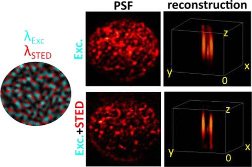
Stimulated emission depletion (STED) microscopy has emerged as a powerful technique providing visualization of biological structures at the molecular level in living samples. In this technique, the diffraction limit is broken by selectively depleting the fluorophore’s excited state by stimulated emission, typically using a donut-shaped optical vortex beam. STED microscopy performs exceptionally well in degraded optical conditions, such as living tissues. Nevertheless, photobleaching and acquisition time are among the main challenges for imaging large volumetric fields of view. In this regard, random light beams such as speckle patterns have proved to be especially promising for three-dimensional imaging in compressed sensing schemes. Taking advantage of the high spatial density of intrinsic optical vortices in speckles─one of the most commonly used types of structured beams in STED microscopy─we propose here a novel scheme that employs speckles for performing STED microscopy. Two speckle patterns are generated at the excitation and the depletion wavelengths, respectively, exhibiting inverted intensity contrasts. We illustrate spatial resolution enhancement using complementary speckles as excitation and depletion beams on both fluorescent beads and biological samples. Our results establish a robust method for super-resolved three-dimensional imaging with promising perspectives in terms of temporal resolution and photobleaching.
期刊介绍:
Published as soon as accepted and summarized in monthly issues, ACS Photonics will publish Research Articles, Letters, Perspectives, and Reviews, to encompass the full scope of published research in this field.


 分享
分享
 求助内容:
求助内容: 应助结果提醒方式:
 扫码关注我们
