Maria Carolina Lopes de Souza Ribeiro, Beatriz Araújo Jacinto Ferreira, Ana Carolina Freitas Ribeiro, Fabiana Mantovani Gomes França, Cecilia Pedroso TURSSi, Roberta Tarkany Basting, Waldemir Francisco Vieira-Junior
{"title":"Occlusion, acid resistance, and elemental characterization of dentin treated with desensitizing agents.","authors":"Maria Carolina Lopes de Souza Ribeiro, Beatriz Araújo Jacinto Ferreira, Ana Carolina Freitas Ribeiro, Fabiana Mantovani Gomes França, Cecilia Pedroso TURSSi, Roberta Tarkany Basting, Waldemir Francisco Vieira-Junior","doi":"10.1590/1807-3107bor-2025.vol39.016","DOIUrl":null,"url":null,"abstract":"<p><p>The objective of this study was to evaluate the occlusion potential of in-office desensitizing agents, and characterize the human dentin elements after acid exposure. Twelve human dentin discs were sectioned into four specimens each, and randomized into treatments (n = 20): no treatment (negative control); no treatment and 6% citric acid exposure (positive control); application of Gluma desensitizer (Heraeus Kulzer) or PRG Barrier Coat (Shofu), followed by 6% citric acid exposure. Occlusion and dentin surface characteristics were determined by scanning electron microscopy (SEM, n = 10), and elemental composition (at%), by energy-dispersive X-ray spectroscopy (EDS, n = 10). Three calibrated, blinded evaluators used SEM to categorize the occlusion potential: 1 = occluded, 2 = partially unoccluded, 3 = equally occluded/unoccluded, 4 = partially occluded, 5 = unoccluded. Data were analyzed by weighted kappa, Friedman, and Nemenyi tests (α = 0.05). For SEM, mean occlusion scores were higher for the PRG Barrier Coat than the positive control (p = 0.0235). Most specimens in the controls scored 4 or 5. The most frequent scores for PRG Barrier Coat were 1(60%) and 2(20%), while 30% of Gluma specimens scored 1 and 2. Gluma showed intratubular precipitation, while PRG Barrier Coat covered dentinal tubules totally or partially. For EDS, the K% was lower for Gluma than the negative control (p = 0.0046), with Si peaks in dentin treated with PRG Barrier Coat. The bioactive in-office desensitizing agent with S-PRG filler (PRG Barrier Coat) promoted dentin tubule occlusion, and persisted after exposure to acid.</p>","PeriodicalId":9240,"journal":{"name":"Brazilian oral research","volume":"39 ","pages":"e016"},"PeriodicalIF":1.3000,"publicationDate":"2025-02-07","publicationTypes":"Journal Article","fieldsOfStudy":null,"isOpenAccess":false,"openAccessPdf":"https://www.ncbi.nlm.nih.gov/pmc/articles/PMC11808701/pdf/","citationCount":"0","resultStr":null,"platform":"Semanticscholar","paperid":null,"PeriodicalName":"Brazilian oral research","FirstCategoryId":"3","ListUrlMain":"https://doi.org/10.1590/1807-3107bor-2025.vol39.016","RegionNum":4,"RegionCategory":"医学","ArticlePicture":[],"TitleCN":null,"AbstractTextCN":null,"PMCID":null,"EPubDate":"2025/1/1 0:00:00","PubModel":"eCollection","JCR":"Q3","JCRName":"DENTISTRY, ORAL SURGERY & MEDICINE","Score":null,"Total":0}
引用次数: 0
Abstract
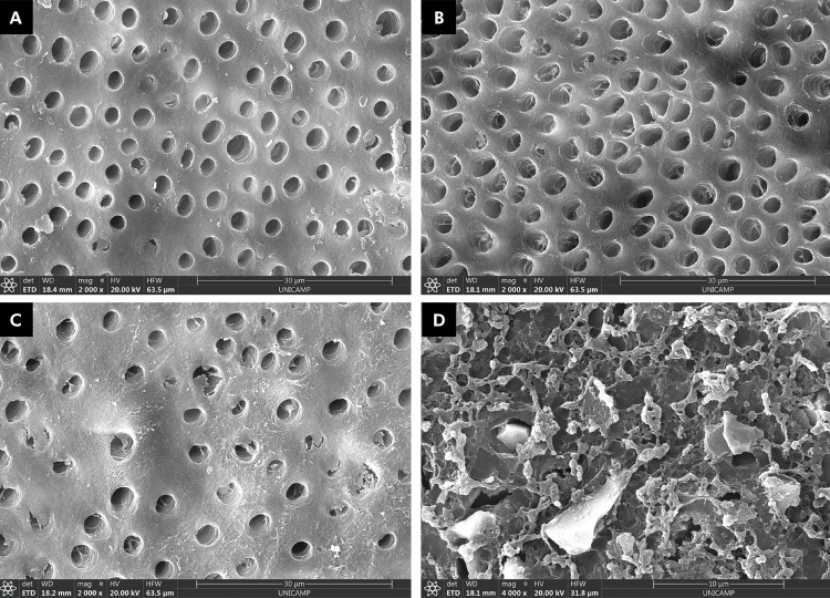
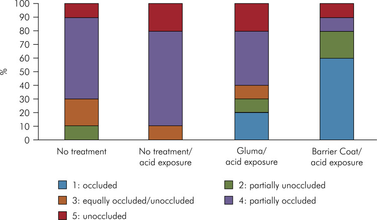
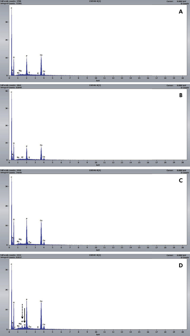
The objective of this study was to evaluate the occlusion potential of in-office desensitizing agents, and characterize the human dentin elements after acid exposure. Twelve human dentin discs were sectioned into four specimens each, and randomized into treatments (n = 20): no treatment (negative control); no treatment and 6% citric acid exposure (positive control); application of Gluma desensitizer (Heraeus Kulzer) or PRG Barrier Coat (Shofu), followed by 6% citric acid exposure. Occlusion and dentin surface characteristics were determined by scanning electron microscopy (SEM, n = 10), and elemental composition (at%), by energy-dispersive X-ray spectroscopy (EDS, n = 10). Three calibrated, blinded evaluators used SEM to categorize the occlusion potential: 1 = occluded, 2 = partially unoccluded, 3 = equally occluded/unoccluded, 4 = partially occluded, 5 = unoccluded. Data were analyzed by weighted kappa, Friedman, and Nemenyi tests (α = 0.05). For SEM, mean occlusion scores were higher for the PRG Barrier Coat than the positive control (p = 0.0235). Most specimens in the controls scored 4 or 5. The most frequent scores for PRG Barrier Coat were 1(60%) and 2(20%), while 30% of Gluma specimens scored 1 and 2. Gluma showed intratubular precipitation, while PRG Barrier Coat covered dentinal tubules totally or partially. For EDS, the K% was lower for Gluma than the negative control (p = 0.0046), with Si peaks in dentin treated with PRG Barrier Coat. The bioactive in-office desensitizing agent with S-PRG filler (PRG Barrier Coat) promoted dentin tubule occlusion, and persisted after exposure to acid.


 分享
分享
 求助内容:
求助内容: 应助结果提醒方式:
 扫码关注我们
