Jon André Ottesen, Tryggve Storas, Svein Are Sirirud Vatnehol, Grethe Løvland, Einar Osland Vik-Mo, Till Schellhorn, Karoline Skogen, Christopher Larsson, Atle Bjørnerud, Inge Rasmus Groote-Eindbaas, Matthan W A Caan
{"title":"Deep learning-based Intraoperative MRI reconstruction.","authors":"Jon André Ottesen, Tryggve Storas, Svein Are Sirirud Vatnehol, Grethe Løvland, Einar Osland Vik-Mo, Till Schellhorn, Karoline Skogen, Christopher Larsson, Atle Bjørnerud, Inge Rasmus Groote-Eindbaas, Matthan W A Caan","doi":"10.1186/s41747-024-00548-9","DOIUrl":null,"url":null,"abstract":"<p><strong>Background: </strong>We retrospectively evaluated the quality of deep learning (DL) reconstructions of on-scanner accelerated intraoperative MRI (iMRI) during respective brain tumor surgery.</p><p><strong>Methods: </strong>Accelerated iMRI was performed using dual surface coils positioned around the area of resection. A DL model was trained on the fastMRI neuro dataset to mimic the data from the iMRI protocol. The evaluation was performed on imaging material from 40 patients imaged from Nov 1, 2021, to June 1, 2023, who underwent iMRI during tumor resection surgery. A comparative analysis was conducted between the conventional compressed sense (CS) method and the trained DL reconstruction method. Blinded evaluation of multiple image quality metrics was performed by two neuroradiologists and one neurosurgeon using a 1-to-5 Likert scale (1, nondiagnostic; 2, poor; 3, acceptable; 4, good; and 5, excellent), and the favored reconstruction variant.</p><p><strong>Results: </strong>The DL reconstruction was strongly favored or favored over the CS reconstruction for 33/40, 39/40, and 8/40 of cases for readers 1, 2, and 3, respectively. For the evaluation metrics, the DL reconstructions had a higher score than their respective CS counterparts for 72%, 72%, and 14% of the cases for readers 1, 2, and 3, respectively. Still, the DL reconstructions exhibited shortcomings such as a striping artifact and reduced signal.</p><p><strong>Conclusion: </strong>DL shows promise in allowing for high-quality reconstructions of iMRI. The neuroradiologists noted an improvement in the perceived spatial resolution, signal-to-noise ratio, diagnostic confidence, diagnostic conspicuity, and spatial resolution compared to CS, while the neurosurgeon preferred the CS reconstructions across all metrics.</p><p><strong>Relevance statement: </strong>DL shows promise to allow for high-quality reconstructions of iMRI, however, due to the challenging setting of iMRI, further optimization is needed.</p><p><strong>Key points: </strong>iMRI is a surgical tool with a challenging image setting. DL allowed for high-quality reconstructions of iMRI. Additional optimization is needed due to the challenging intraoperative setting.</p>","PeriodicalId":36926,"journal":{"name":"European Radiology Experimental","volume":"9 1","pages":"29"},"PeriodicalIF":3.6000,"publicationDate":"2025-02-25","publicationTypes":"Journal Article","fieldsOfStudy":null,"isOpenAccess":false,"openAccessPdf":"https://www.ncbi.nlm.nih.gov/pmc/articles/PMC11861787/pdf/","citationCount":"0","resultStr":null,"platform":"Semanticscholar","paperid":null,"PeriodicalName":"European Radiology Experimental","FirstCategoryId":"1085","ListUrlMain":"https://doi.org/10.1186/s41747-024-00548-9","RegionNum":0,"RegionCategory":null,"ArticlePicture":[],"TitleCN":null,"AbstractTextCN":null,"PMCID":null,"EPubDate":"","PubModel":"","JCR":"Q1","JCRName":"RADIOLOGY, NUCLEAR MEDICINE & MEDICAL IMAGING","Score":null,"Total":0}
引用次数: 0
Abstract
Background: We retrospectively evaluated the quality of deep learning (DL) reconstructions of on-scanner accelerated intraoperative MRI (iMRI) during respective brain tumor surgery.
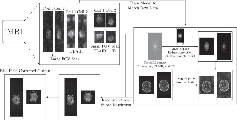
Methods: Accelerated iMRI was performed using dual surface coils positioned around the area of resection. A DL model was trained on the fastMRI neuro dataset to mimic the data from the iMRI protocol. The evaluation was performed on imaging material from 40 patients imaged from Nov 1, 2021, to June 1, 2023, who underwent iMRI during tumor resection surgery. A comparative analysis was conducted between the conventional compressed sense (CS) method and the trained DL reconstruction method. Blinded evaluation of multiple image quality metrics was performed by two neuroradiologists and one neurosurgeon using a 1-to-5 Likert scale (1, nondiagnostic; 2, poor; 3, acceptable; 4, good; and 5, excellent), and the favored reconstruction variant.
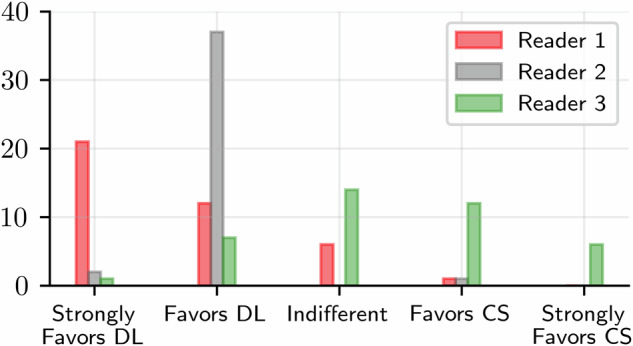
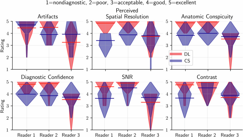
Results: The DL reconstruction was strongly favored or favored over the CS reconstruction for 33/40, 39/40, and 8/40 of cases for readers 1, 2, and 3, respectively. For the evaluation metrics, the DL reconstructions had a higher score than their respective CS counterparts for 72%, 72%, and 14% of the cases for readers 1, 2, and 3, respectively. Still, the DL reconstructions exhibited shortcomings such as a striping artifact and reduced signal.
Conclusion: DL shows promise in allowing for high-quality reconstructions of iMRI. The neuroradiologists noted an improvement in the perceived spatial resolution, signal-to-noise ratio, diagnostic confidence, diagnostic conspicuity, and spatial resolution compared to CS, while the neurosurgeon preferred the CS reconstructions across all metrics.
Relevance statement: DL shows promise to allow for high-quality reconstructions of iMRI, however, due to the challenging setting of iMRI, further optimization is needed.
Key points: iMRI is a surgical tool with a challenging image setting. DL allowed for high-quality reconstructions of iMRI. Additional optimization is needed due to the challenging intraoperative setting.


 分享
分享
 求助内容:
求助内容: 应助结果提醒方式:
 扫码关注我们
