{"title":"A CT-based interpretable deep learning signature for predicting PD-L1 expression in bladder cancer: a two-center study.","authors":"Xiaomeng Han, Jing Guan, Li Guo, Qiyan Jiao, Kexin Wang, Feng Hou, Shunli Liu, Shifeng Yang, Chencui Huang, Wenbin Cong, Hexiang Wang","doi":"10.1186/s40644-025-00849-1","DOIUrl":null,"url":null,"abstract":"<p><strong>Background: </strong>To construct and assess a deep learning (DL) signature that employs computed tomography imaging to predict the expression status of programmed cell death ligand 1 in patients with bladder cancer (BCa).</p><p><strong>Methods: </strong>This retrospective study included 190 patients from two hospitals who underwent surgical removal of BCa (training set/external validation set, 127/63). We used convolutional neural network and radiomics machine learning technology to generate prediction models. We then compared the performance of the DL signature with the radiomics machine learning signature and selected the optimal signature to build a nomogram with the clinical model. Finally, the internal forecasting process of the DL signature was explained using Shapley additive explanation technology.</p><p><strong>Results: </strong>On the external validation set, the DL signature had an area under the curve of 0.857 (95% confidence interval: 0.745-0.932), and demonstrated superior prediction performance in comparison with the other models. SHAP expression images revealed that the prediction of PD-L1 expression status is mainly influenced by the tumor edge region, particularly the area close to the bladder wall.</p><p><strong>Conclusions: </strong>The DL signature performed well in comparison with other models and proved to be a valuable, dependable, and interpretable tool for predicting programmed cell death ligand 1 expression status in patients with BCa.</p>","PeriodicalId":9548,"journal":{"name":"Cancer Imaging","volume":"25 1","pages":"27"},"PeriodicalIF":3.5000,"publicationDate":"2025-03-10","publicationTypes":"Journal Article","fieldsOfStudy":null,"isOpenAccess":false,"openAccessPdf":"https://www.ncbi.nlm.nih.gov/pmc/articles/PMC11892212/pdf/","citationCount":"0","resultStr":null,"platform":"Semanticscholar","paperid":null,"PeriodicalName":"Cancer Imaging","FirstCategoryId":"3","ListUrlMain":"https://doi.org/10.1186/s40644-025-00849-1","RegionNum":2,"RegionCategory":"医学","ArticlePicture":[],"TitleCN":null,"AbstractTextCN":null,"PMCID":null,"EPubDate":"","PubModel":"","JCR":"Q2","JCRName":"ONCOLOGY","Score":null,"Total":0}
引用次数: 0
Abstract
Background: To construct and assess a deep learning (DL) signature that employs computed tomography imaging to predict the expression status of programmed cell death ligand 1 in patients with bladder cancer (BCa).
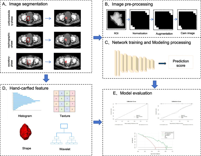
Methods: This retrospective study included 190 patients from two hospitals who underwent surgical removal of BCa (training set/external validation set, 127/63). We used convolutional neural network and radiomics machine learning technology to generate prediction models. We then compared the performance of the DL signature with the radiomics machine learning signature and selected the optimal signature to build a nomogram with the clinical model. Finally, the internal forecasting process of the DL signature was explained using Shapley additive explanation technology.
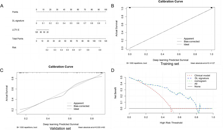
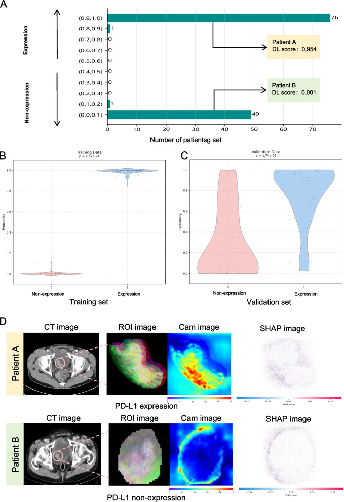
Results: On the external validation set, the DL signature had an area under the curve of 0.857 (95% confidence interval: 0.745-0.932), and demonstrated superior prediction performance in comparison with the other models. SHAP expression images revealed that the prediction of PD-L1 expression status is mainly influenced by the tumor edge region, particularly the area close to the bladder wall.
Conclusions: The DL signature performed well in comparison with other models and proved to be a valuable, dependable, and interpretable tool for predicting programmed cell death ligand 1 expression status in patients with BCa.
Cancer ImagingONCOLOGY-RADIOLOGY, NUCLEAR MEDICINE & MEDICAL IMAGING
CiteScore
7.00
自引率
0.00%
发文量
66
审稿时长
>12 weeks
期刊介绍:
Cancer Imaging is an open access, peer-reviewed journal publishing original articles, reviews and editorials written by expert international radiologists working in oncology.
The journal encompasses CT, MR, PET, ultrasound, radionuclide and multimodal imaging in all kinds of malignant tumours, plus new developments, techniques and innovations. Topics of interest include:
Breast Imaging
Chest
Complications of treatment
Ear, Nose & Throat
Gastrointestinal
Hepatobiliary & Pancreatic
Imaging biomarkers
Interventional
Lymphoma
Measurement of tumour response
Molecular functional imaging
Musculoskeletal
Neuro oncology
Nuclear Medicine
Paediatric.


 分享
分享
 求助内容:
求助内容: 应助结果提醒方式:
 扫码关注我们
