Jitender Jitender, Teresa Hong, Anakim Sherman, Patty Wong, Eric Aniogo, Maciej Kujawski, John E Shively, Paul J Yazaki
{"title":"Development of Anti-CEA C<sub>H</sub>2 Domain-Deleted Antibody (M5A∆C<sub>H</sub>2) for the PET Imaging of Colorectal Cancer.","authors":"Jitender Jitender, Teresa Hong, Anakim Sherman, Patty Wong, Eric Aniogo, Maciej Kujawski, John E Shively, Paul J Yazaki","doi":"10.1007/s11307-025-01997-3","DOIUrl":null,"url":null,"abstract":"<p><strong>Purpose: </strong>Recombinant antibody fragments represent a novel class of in vivo biological immunoPET imaging agents. This study developed a series of anti-carcinoembryonic antigen (CEA) C<sub>H</sub>2 domain-deleted antibodies to evaluate their rapid, high-level tumor targeting combined with fast blood clearance for immunoPET imaging in two colorectal cancer mouse models.</p><p><strong>Procedure: </strong>A series of humanized anti-CEA M5A∆C<sub>H</sub>2 recombinant antibody fragments were synthesized via transient mammalian expression and purified using a two-step process. The M5A∆CH2 antibody series was characterized by HPLC-SEC, SDS-PAGE and binding affinities. The M5A∆C<sub>H</sub>2-C5 antibody, which has five disulfide bridges in the modified IgG1/IgG3 hinge domain, was selected for positron emission tomography (PET) imaging. Site-specific thiol conjugation of the reduced hinge disulfides with the 1,4,7,10 tetraazacyclododecane-1,4,7-triacetic acid trisodium salt-vinyl sulfone (DO3A-VS) chelate was performed, followed by labeling with [<sup>64</sup>Cu-CuCl<sub>2</sub>]. The [<sup>64</sup>Cu]Cu-DO3A-M5A∆C<sub>H</sub>2-C5 was evaluated for CEA-positive tumor PET imaging at serial time points, pharmacokinetics and a terminal biodistribution study conducted in two CEA-positive colorectal cancer mouse models.</p><p><strong>Results: </strong>The anti-CEA M5A∆C<sub>H</sub>2 antibodies had high expression, were purified using a new CH3 domain affinity resin and were stable up to one year. ImmunoPET imaging and biodistribution studies were performed in athymic mice bearing human colorectal cancer LS174T tumors and immunocompetent transgenic-CEA (Tg-CEA) mice bearing MC-38 tumors transfected with the human CEA gene. The [<sup>64</sup>Cu]Cu-DO3A-M5A∆C<sub>H</sub>2-C5 showed rapid, high tumor localization and the expected fast blood clearance.</p><p><strong>Conclusions: </strong>A series of humanized anti-CEA M5A∆C<sub>H</sub>2 antibodies were designed for immunoPET imaging of colorectal cancer, and the [<sup>64</sup>Cu]Cu-DO3A-M5A∆C<sub>H</sub>2-C5 showed high tumor targeting and fast blood clearance supporting its potential for clinical trials.</p>","PeriodicalId":18760,"journal":{"name":"Molecular Imaging and Biology","volume":" ","pages":"192-200"},"PeriodicalIF":2.5000,"publicationDate":"2025-04-01","publicationTypes":"Journal Article","fieldsOfStudy":null,"isOpenAccess":false,"openAccessPdf":"https://www.ncbi.nlm.nih.gov/pmc/articles/PMC12062029/pdf/","citationCount":"0","resultStr":null,"platform":"Semanticscholar","paperid":null,"PeriodicalName":"Molecular Imaging and Biology","FirstCategoryId":"3","ListUrlMain":"https://doi.org/10.1007/s11307-025-01997-3","RegionNum":4,"RegionCategory":"医学","ArticlePicture":[],"TitleCN":null,"AbstractTextCN":null,"PMCID":null,"EPubDate":"2025/3/14 0:00:00","PubModel":"Epub","JCR":"Q2","JCRName":"RADIOLOGY, NUCLEAR MEDICINE & MEDICAL IMAGING","Score":null,"Total":0}
引用次数: 0
Abstract
Purpose: Recombinant antibody fragments represent a novel class of in vivo biological immunoPET imaging agents. This study developed a series of anti-carcinoembryonic antigen (CEA) CH2 domain-deleted antibodies to evaluate their rapid, high-level tumor targeting combined with fast blood clearance for immunoPET imaging in two colorectal cancer mouse models.
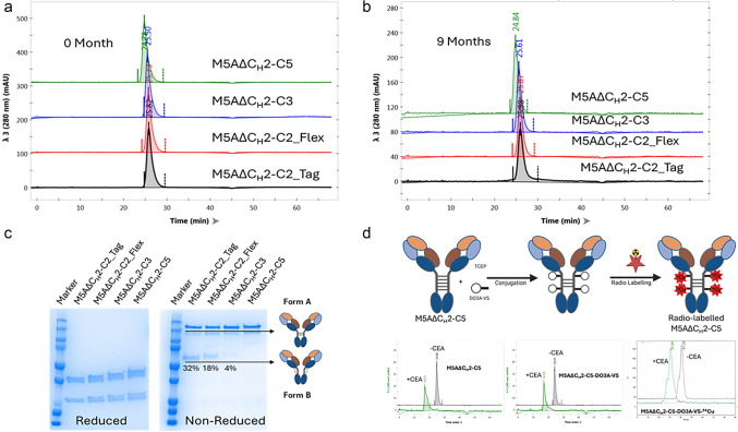
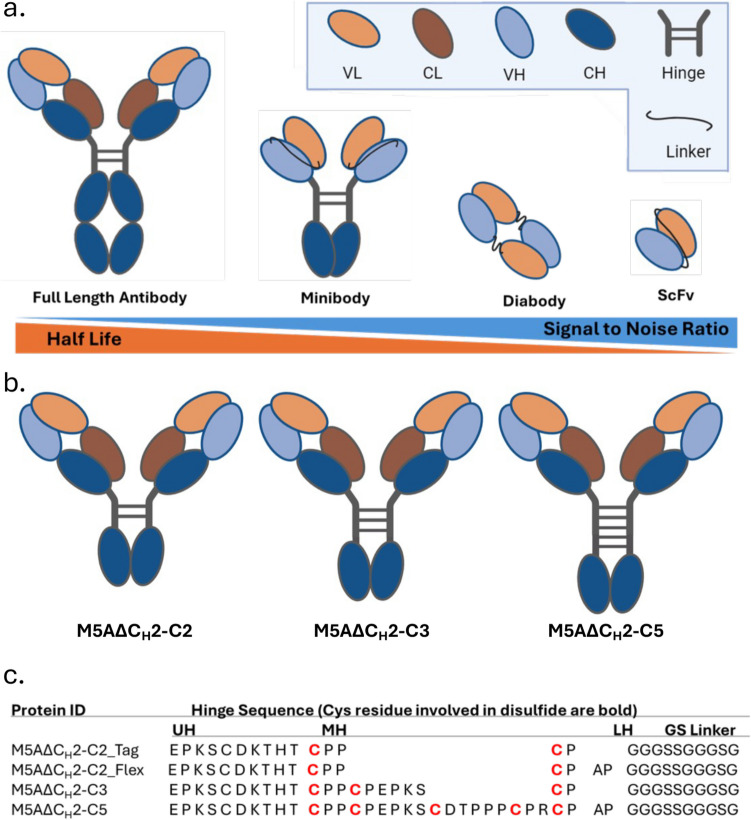
Procedure: A series of humanized anti-CEA M5A∆CH2 recombinant antibody fragments were synthesized via transient mammalian expression and purified using a two-step process. The M5A∆CH2 antibody series was characterized by HPLC-SEC, SDS-PAGE and binding affinities. The M5A∆CH2-C5 antibody, which has five disulfide bridges in the modified IgG1/IgG3 hinge domain, was selected for positron emission tomography (PET) imaging. Site-specific thiol conjugation of the reduced hinge disulfides with the 1,4,7,10 tetraazacyclododecane-1,4,7-triacetic acid trisodium salt-vinyl sulfone (DO3A-VS) chelate was performed, followed by labeling with [64Cu-CuCl2]. The [64Cu]Cu-DO3A-M5A∆CH2-C5 was evaluated for CEA-positive tumor PET imaging at serial time points, pharmacokinetics and a terminal biodistribution study conducted in two CEA-positive colorectal cancer mouse models.
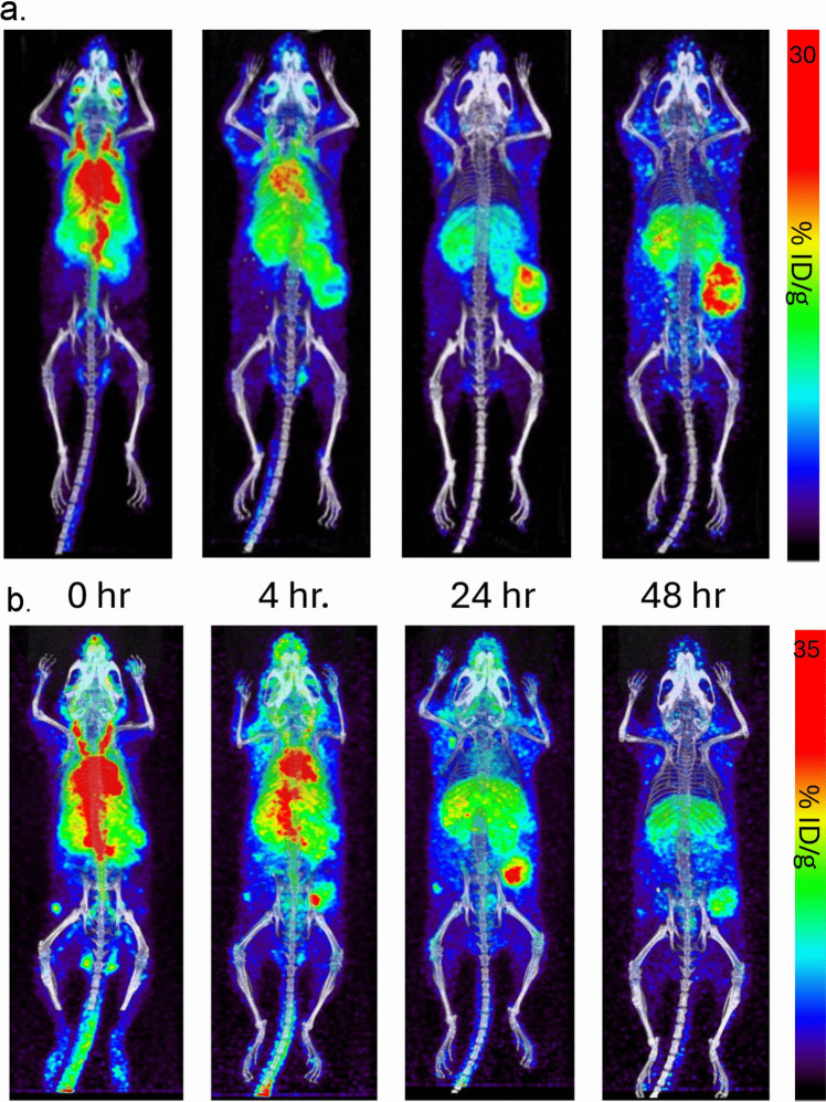
Results: The anti-CEA M5A∆CH2 antibodies had high expression, were purified using a new CH3 domain affinity resin and were stable up to one year. ImmunoPET imaging and biodistribution studies were performed in athymic mice bearing human colorectal cancer LS174T tumors and immunocompetent transgenic-CEA (Tg-CEA) mice bearing MC-38 tumors transfected with the human CEA gene. The [64Cu]Cu-DO3A-M5A∆CH2-C5 showed rapid, high tumor localization and the expected fast blood clearance.
Conclusions: A series of humanized anti-CEA M5A∆CH2 antibodies were designed for immunoPET imaging of colorectal cancer, and the [64Cu]Cu-DO3A-M5A∆CH2-C5 showed high tumor targeting and fast blood clearance supporting its potential for clinical trials.
期刊介绍:
Molecular Imaging and Biology (MIB) invites original contributions (research articles, review articles, commentaries, etc.) on the utilization of molecular imaging (i.e., nuclear imaging, optical imaging, autoradiography and pathology, MRI, MPI, ultrasound imaging, radiomics/genomics etc.) to investigate questions related to biology and health. The objective of MIB is to provide a forum to the discovery of molecular mechanisms of disease through the use of imaging techniques. We aim to investigate the biological nature of disease in patients and establish new molecular imaging diagnostic and therapy procedures.
Some areas that are covered are:
Preclinical and clinical imaging of macromolecular targets (e.g., genes, receptors, enzymes) involved in significant biological processes.
The design, characterization, and study of new molecular imaging probes and contrast agents for the functional interrogation of macromolecular targets.
Development and evaluation of imaging systems including instrumentation, image reconstruction algorithms, image analysis, and display.
Development of molecular assay approaches leading to quantification of the biological information obtained in molecular imaging.
Study of in vivo animal models of disease for the development of new molecular diagnostics and therapeutics.
Extension of in vitro and in vivo discoveries using disease models, into well designed clinical research investigations.
Clinical molecular imaging involving clinical investigations, clinical trials and medical management or cost-effectiveness studies.


 分享
分享

 求助内容:
求助内容: 应助结果提醒方式:
 扫码关注我们
