Christian Happel, Larissa Völler, Benjamin Bockisch, Daniel Groener, Britta Leonhäuser, Frank Grünwald, Amir Sabet
{"title":"Development of a CT-less SPECT Acquisition Protocol for Kidney Dosimetry in <sup>177</sup>Lu-PSMA-617 Radioligand Therapy.","authors":"Christian Happel, Larissa Völler, Benjamin Bockisch, Daniel Groener, Britta Leonhäuser, Frank Grünwald, Amir Sabet","doi":"10.1007/s11307-025-01998-2","DOIUrl":null,"url":null,"abstract":"<p><strong>Purpose: </strong>Targeted radioligand therapy of metastatic castration-resistant prostate cancer (mCRPC) with <sup>177</sup>Lu-PSMA (RLT) requires sufficient dose monitoring of the kidneys. Currently, dosimetry using SPECT/CT-imaging is the most preferred method. However, SPECT/CT is a time-consuming procedure and comprises additional radiation exposure to the patient. Moreover, not every therapeutic nuclear medicine facility has access to SPECT/CT. Therefore, the aim of this study was to develop a new procedure of kidney dosimetry without the use of SPECT/CT and evaluate this method in a large cohort of patients with mCRPC undergoing RLT.</p><p><strong>Procedures: </strong>A dedicated torso phantom with kidneys filled with a solution of <sup>177</sup>Lu-PSMA was used for quantitative calibration of a SPECT-camera. The calculated sensitivity was adapted according to the individual attenuation of the patient in four directions from the kidney surface to the body surface (ventral, dorsal, left and right) obtained from a previously performed CT. A total of 196 patients undergoing 926 cycles of <sup>177</sup>Lu-PSMA therapy were retrospectively analyzed. Abdominal SPECT was performed 24, 48 and 72 h after administration of <sup>177</sup>Lu-PSMA including scatter and dead-time correction in every patient. Kidney dose was calculated using an individual attenuation-based procedure and compared to values from international literature.</p><p><strong>Results: </strong>Volumes of interest of the kidneys were drawn in the three sequential SPECT-images to calculate intra-renal effective half-life. Absolute quantification of activity in the kidneys was accomplished obtaining a patient individual sensitivity based on the individual attenuation in the patient. Kidney dose was then calculated applying a bi-exponential time activity curve in Microsoft EXCEL. Mean kidney dose per administered activity was 0.54 (± 0.26) Gy/GBq.</p><p><strong>Conclusions: </strong>With the presented procedure a reliable kidney dosimetry is possible without the use of SPECT/CT. Facilities without SPECT/CT are therefore able to perform an adequate kidney dosimetry without additional radiation exposure for the patient.</p>","PeriodicalId":18760,"journal":{"name":"Molecular Imaging and Biology","volume":" ","pages":"400-409"},"PeriodicalIF":2.5000,"publicationDate":"2025-06-01","publicationTypes":"Journal Article","fieldsOfStudy":null,"isOpenAccess":false,"openAccessPdf":"https://www.ncbi.nlm.nih.gov/pmc/articles/PMC12162791/pdf/","citationCount":"0","resultStr":null,"platform":"Semanticscholar","paperid":null,"PeriodicalName":"Molecular Imaging and Biology","FirstCategoryId":"3","ListUrlMain":"https://doi.org/10.1007/s11307-025-01998-2","RegionNum":4,"RegionCategory":"医学","ArticlePicture":[],"TitleCN":null,"AbstractTextCN":null,"PMCID":null,"EPubDate":"2025/3/20 0:00:00","PubModel":"Epub","JCR":"Q2","JCRName":"RADIOLOGY, NUCLEAR MEDICINE & MEDICAL IMAGING","Score":null,"Total":0}
引用次数: 0
Abstract
Purpose: Targeted radioligand therapy of metastatic castration-resistant prostate cancer (mCRPC) with 177Lu-PSMA (RLT) requires sufficient dose monitoring of the kidneys. Currently, dosimetry using SPECT/CT-imaging is the most preferred method. However, SPECT/CT is a time-consuming procedure and comprises additional radiation exposure to the patient. Moreover, not every therapeutic nuclear medicine facility has access to SPECT/CT. Therefore, the aim of this study was to develop a new procedure of kidney dosimetry without the use of SPECT/CT and evaluate this method in a large cohort of patients with mCRPC undergoing RLT.
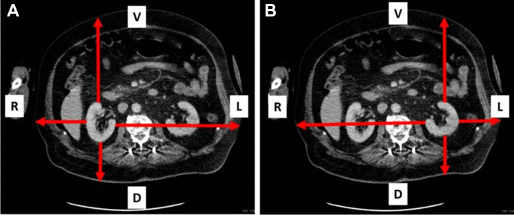
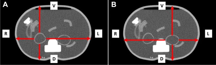
Procedures: A dedicated torso phantom with kidneys filled with a solution of 177Lu-PSMA was used for quantitative calibration of a SPECT-camera. The calculated sensitivity was adapted according to the individual attenuation of the patient in four directions from the kidney surface to the body surface (ventral, dorsal, left and right) obtained from a previously performed CT. A total of 196 patients undergoing 926 cycles of 177Lu-PSMA therapy were retrospectively analyzed. Abdominal SPECT was performed 24, 48 and 72 h after administration of 177Lu-PSMA including scatter and dead-time correction in every patient. Kidney dose was calculated using an individual attenuation-based procedure and compared to values from international literature.
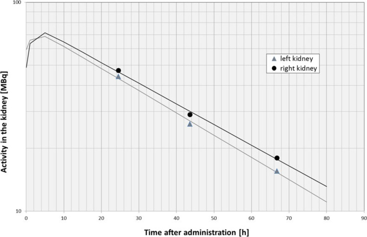
Results: Volumes of interest of the kidneys were drawn in the three sequential SPECT-images to calculate intra-renal effective half-life. Absolute quantification of activity in the kidneys was accomplished obtaining a patient individual sensitivity based on the individual attenuation in the patient. Kidney dose was then calculated applying a bi-exponential time activity curve in Microsoft EXCEL. Mean kidney dose per administered activity was 0.54 (± 0.26) Gy/GBq.
Conclusions: With the presented procedure a reliable kidney dosimetry is possible without the use of SPECT/CT. Facilities without SPECT/CT are therefore able to perform an adequate kidney dosimetry without additional radiation exposure for the patient.
期刊介绍:
Molecular Imaging and Biology (MIB) invites original contributions (research articles, review articles, commentaries, etc.) on the utilization of molecular imaging (i.e., nuclear imaging, optical imaging, autoradiography and pathology, MRI, MPI, ultrasound imaging, radiomics/genomics etc.) to investigate questions related to biology and health. The objective of MIB is to provide a forum to the discovery of molecular mechanisms of disease through the use of imaging techniques. We aim to investigate the biological nature of disease in patients and establish new molecular imaging diagnostic and therapy procedures.
Some areas that are covered are:
Preclinical and clinical imaging of macromolecular targets (e.g., genes, receptors, enzymes) involved in significant biological processes.
The design, characterization, and study of new molecular imaging probes and contrast agents for the functional interrogation of macromolecular targets.
Development and evaluation of imaging systems including instrumentation, image reconstruction algorithms, image analysis, and display.
Development of molecular assay approaches leading to quantification of the biological information obtained in molecular imaging.
Study of in vivo animal models of disease for the development of new molecular diagnostics and therapeutics.
Extension of in vitro and in vivo discoveries using disease models, into well designed clinical research investigations.
Clinical molecular imaging involving clinical investigations, clinical trials and medical management or cost-effectiveness studies.


 分享
分享

 求助内容:
求助内容: 应助结果提醒方式:
 扫码关注我们
