Arnaud Dieudonné, Aya Terro, Arthur Dumouchel, Solène Perret, Agathe Edet-Sanson, Pierre Vera, Sébastien Hapdey, Romain Modzelewski, David Tonnelet, Pierre Decazes
{"title":"Towards fully automatized [177Lu]Lu-PSMA personalized dosimetry based on 360° CZT whole-body SPECT/CT: a proof-of-concept.","authors":"Arnaud Dieudonné, Aya Terro, Arthur Dumouchel, Solène Perret, Agathe Edet-Sanson, Pierre Vera, Sébastien Hapdey, Romain Modzelewski, David Tonnelet, Pierre Decazes","doi":"10.1186/s40658-025-00727-6","DOIUrl":null,"url":null,"abstract":"<p><strong>Background: </strong>The advent of 360° CZT gamma-cameras allows to conceive personalised dosimetry studies from whole-body SPECT/CT data. We aimed to demonstrate the proof-of-concept of an automated personalized dosimetry pipeline for [<sup>177</sup>Lu]Lu-PSMA organ dosimetry, called SimpleDose, and to compare to other dosimetry approaches.</p><p><strong>Methods: </strong>The organ segmentation is based on a nnU-Net framework that was trained to allow for the segmentation of 23 organs and structures over all the body. The method implemented to model the energy deposition is the collapsed-cone-superposition (CCS) taking into account non-uniform activity and density distributions. Ten patients with metastatic castration resistant prostate cancer treated [<sup>177</sup>Lu]Lu-PSMA-617 were included. All SPECT/CT acquisitions were performed on a VERITON-CT 200 (Spectrum Dynamics®, Caesarea, Israel) from head to mid-thigh with 5 min per bed. The absorbed-dose-rates were computed with SimpleDose and compared with organ-level MIRD approach and local-deposition-method (LDM) for bone marrow, kidneys, liver, lungs, pancreas, salivary glands and spleen. Finally, an example of multi-time-point and single-time-point dosimetry is given.</p><p><strong>Results: </strong>The median (IQR) calculation time with SimpleDose (SD), for segmentation, computation of dose-rates and descriptive statistics was 161 (23) seconds at a resolution of 2.46 × 2.46 × 2.46 mm<sup>3</sup> (Intel Xeon 20 × 3.70 GHz CPU computer). The median (IQR) differences between SD and MIRD and LDM, were respectively 1.8 (61) % and - 16 (76) % in bone marrow, 2.4 (1.5) % and - 93.1 (0.4) % in kidneys, 2.9 (3.4) % and - 9.2 (3.0) % in liver, 21 (13) % and 13 (13) % in lungs, 11 (3.3) % and - 11 (3.0) % in pancreas, 1.1 (12) % and 3.8 (8.4) % in salivary glands, 4.0 (4.3) % and - 10.0 (4.5) % in spleen. For the clinical example, the multi-time-point dosimetry with 4 time-points took 14 min, while the single-time-point approach took 3.5 min from day 1 dataset and 3.3 min from day 3.</p><p><strong>Conclusion: </strong>The SimpleDose platform demonstrated its capability to compute organ-absorbed-dose rates in a simple and fast manner with close results to the standard MIRD approach for soft-tissues organs. SimpleDose is freely available for demonstration purpose as a Software as a Service (SaaS) at https://oncometer3d.com .</p>","PeriodicalId":11559,"journal":{"name":"EJNMMI Physics","volume":"12 1","pages":"25"},"PeriodicalIF":3.2000,"publicationDate":"2025-03-20","publicationTypes":"Journal Article","fieldsOfStudy":null,"isOpenAccess":false,"openAccessPdf":"https://www.ncbi.nlm.nih.gov/pmc/articles/PMC11925831/pdf/","citationCount":"0","resultStr":null,"platform":"Semanticscholar","paperid":null,"PeriodicalName":"EJNMMI Physics","FirstCategoryId":"3","ListUrlMain":"https://doi.org/10.1186/s40658-025-00727-6","RegionNum":2,"RegionCategory":"医学","ArticlePicture":[],"TitleCN":null,"AbstractTextCN":null,"PMCID":null,"EPubDate":"","PubModel":"","JCR":"Q2","JCRName":"RADIOLOGY, NUCLEAR MEDICINE & MEDICAL IMAGING","Score":null,"Total":0}
引用次数: 0
Abstract
Background: The advent of 360° CZT gamma-cameras allows to conceive personalised dosimetry studies from whole-body SPECT/CT data. We aimed to demonstrate the proof-of-concept of an automated personalized dosimetry pipeline for [177Lu]Lu-PSMA organ dosimetry, called SimpleDose, and to compare to other dosimetry approaches.
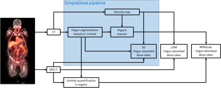
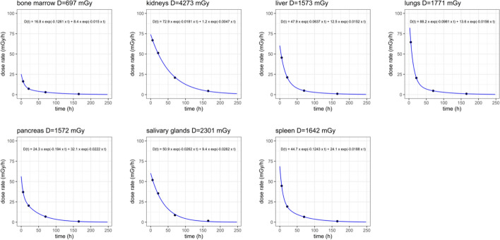
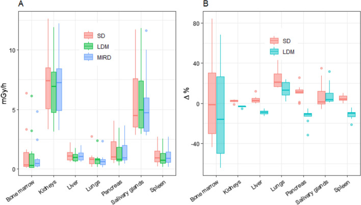
Methods: The organ segmentation is based on a nnU-Net framework that was trained to allow for the segmentation of 23 organs and structures over all the body. The method implemented to model the energy deposition is the collapsed-cone-superposition (CCS) taking into account non-uniform activity and density distributions. Ten patients with metastatic castration resistant prostate cancer treated [177Lu]Lu-PSMA-617 were included. All SPECT/CT acquisitions were performed on a VERITON-CT 200 (Spectrum Dynamics®, Caesarea, Israel) from head to mid-thigh with 5 min per bed. The absorbed-dose-rates were computed with SimpleDose and compared with organ-level MIRD approach and local-deposition-method (LDM) for bone marrow, kidneys, liver, lungs, pancreas, salivary glands and spleen. Finally, an example of multi-time-point and single-time-point dosimetry is given.
Results: The median (IQR) calculation time with SimpleDose (SD), for segmentation, computation of dose-rates and descriptive statistics was 161 (23) seconds at a resolution of 2.46 × 2.46 × 2.46 mm3 (Intel Xeon 20 × 3.70 GHz CPU computer). The median (IQR) differences between SD and MIRD and LDM, were respectively 1.8 (61) % and - 16 (76) % in bone marrow, 2.4 (1.5) % and - 93.1 (0.4) % in kidneys, 2.9 (3.4) % and - 9.2 (3.0) % in liver, 21 (13) % and 13 (13) % in lungs, 11 (3.3) % and - 11 (3.0) % in pancreas, 1.1 (12) % and 3.8 (8.4) % in salivary glands, 4.0 (4.3) % and - 10.0 (4.5) % in spleen. For the clinical example, the multi-time-point dosimetry with 4 time-points took 14 min, while the single-time-point approach took 3.5 min from day 1 dataset and 3.3 min from day 3.
Conclusion: The SimpleDose platform demonstrated its capability to compute organ-absorbed-dose rates in a simple and fast manner with close results to the standard MIRD approach for soft-tissues organs. SimpleDose is freely available for demonstration purpose as a Software as a Service (SaaS) at https://oncometer3d.com .
期刊介绍:
EJNMMI Physics is an international platform for scientists, users and adopters of nuclear medicine with a particular interest in physics matters. As a companion journal to the European Journal of Nuclear Medicine and Molecular Imaging, this journal has a multi-disciplinary approach and welcomes original materials and studies with a focus on applied physics and mathematics as well as imaging systems engineering and prototyping in nuclear medicine. This includes physics-driven approaches or algorithms supported by physics that foster early clinical adoption of nuclear medicine imaging and therapy.


 分享
分享
 求助内容:
求助内容: 应助结果提醒方式:
 扫码关注我们
