Silvia Marchesi, Elin Lundström, Elin Lindström, Jonas Ödmark, Mark Lubberink, Håkan Ahlström, Miklós Lipcsey
{"title":"Enhanced glomerular thrombosis in pronated animals with ARDS.","authors":"Silvia Marchesi, Elin Lundström, Elin Lindström, Jonas Ödmark, Mark Lubberink, Håkan Ahlström, Miklós Lipcsey","doi":"10.1186/s40635-025-00747-7","DOIUrl":null,"url":null,"abstract":"<p><strong>Background: </strong>Prone positioning is part of the management of acute respiratory distress syndrome (ARDS) and has been demonstrated to successfully improve the ventilation-perfusion match and reduce mortality in patients with severe respiratory failure. However, the effect of pronation on other organs than the lungs has not been widely studied. This study aimed to compare abdominal edema, perfusion and inflammation in supine and prone positioning in a porcine ARDS model.</p><p><strong>Methods: </strong>Seventeen piglets were randomized into two groups: a supine group (n = 9) and a prone group (n = 8). Both groups received endotoxemic infusion and were observed for 6 h. Three animals per group underwent positron emission tomography-magnetic resonance imaging (PET-MRI) for imaging acquisition. Hemodynamic and respiratory parameters were recorded throughout the protocol. Inflammation was assessed by measuring cytokine concentrations in blood, ascites and the abdominal organs' tissue. The edema in abdominal organs was assessed by wet-dry ratio and pathophysiological analysis of tissue samples and by MRI and PET measurements from volumes of interest (VOIs) delineated in abdominal organ in MRI and PET images. The abdominal organs' perfusion was also assessed by MRI and PET measurements.</p><p><strong>Results: </strong>The prone group had a faster CO<sub>2</sub> washout and needed a lower positive end-expiratory pressure to maintain the desired oxygenation. In the prone group duodenal edema was lower (measured with wet-dry ratio) and renal perfusion, by both MRI and PET measurements, was lower than half compared to the supine group (MRI, perfusion fraction, f: supine group 0.13; prone group 0.03; p-value 0.002. PET Flow: supine group 1.7; prone group 0.4 ml/cm<sup>3</sup>/min; p-value 0.002). In addition, the histopathological samples of the kidneys showed a higher incidence and extent of glomerular thrombosis in the prone group.</p><p><strong>Conclusions: </strong>In a porcine ARDS model, prone positioning was associated with enhanced glomerular thrombosis and low renal perfusion.</p>","PeriodicalId":13750,"journal":{"name":"Intensive Care Medicine Experimental","volume":"13 1","pages":"36"},"PeriodicalIF":2.8000,"publicationDate":"2025-03-20","publicationTypes":"Journal Article","fieldsOfStudy":null,"isOpenAccess":false,"openAccessPdf":"https://www.ncbi.nlm.nih.gov/pmc/articles/PMC11926287/pdf/","citationCount":"0","resultStr":null,"platform":"Semanticscholar","paperid":null,"PeriodicalName":"Intensive Care Medicine Experimental","FirstCategoryId":"1085","ListUrlMain":"https://doi.org/10.1186/s40635-025-00747-7","RegionNum":0,"RegionCategory":null,"ArticlePicture":[],"TitleCN":null,"AbstractTextCN":null,"PMCID":null,"EPubDate":"","PubModel":"","JCR":"Q2","JCRName":"CRITICAL CARE MEDICINE","Score":null,"Total":0}
引用次数: 0
Abstract
Background: Prone positioning is part of the management of acute respiratory distress syndrome (ARDS) and has been demonstrated to successfully improve the ventilation-perfusion match and reduce mortality in patients with severe respiratory failure. However, the effect of pronation on other organs than the lungs has not been widely studied. This study aimed to compare abdominal edema, perfusion and inflammation in supine and prone positioning in a porcine ARDS model.
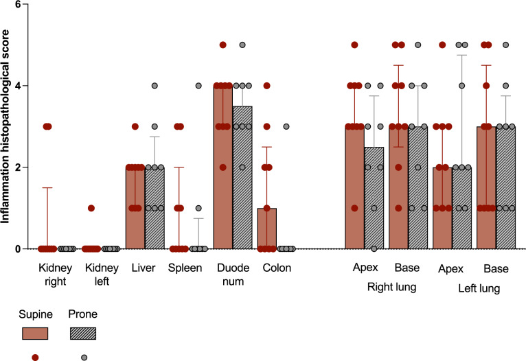
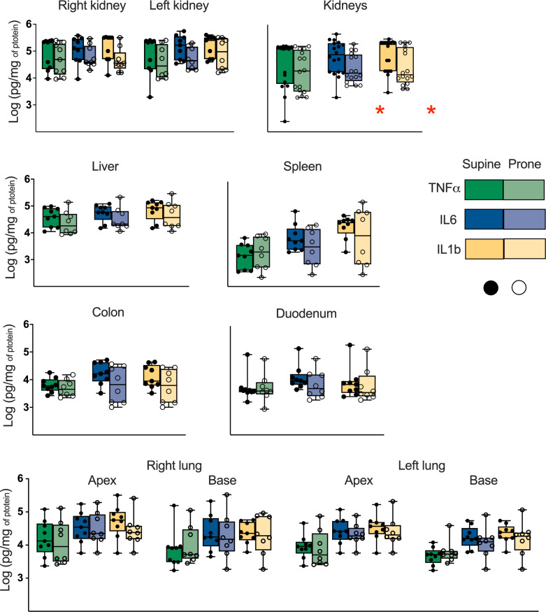
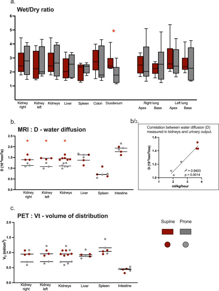
Methods: Seventeen piglets were randomized into two groups: a supine group (n = 9) and a prone group (n = 8). Both groups received endotoxemic infusion and were observed for 6 h. Three animals per group underwent positron emission tomography-magnetic resonance imaging (PET-MRI) for imaging acquisition. Hemodynamic and respiratory parameters were recorded throughout the protocol. Inflammation was assessed by measuring cytokine concentrations in blood, ascites and the abdominal organs' tissue. The edema in abdominal organs was assessed by wet-dry ratio and pathophysiological analysis of tissue samples and by MRI and PET measurements from volumes of interest (VOIs) delineated in abdominal organ in MRI and PET images. The abdominal organs' perfusion was also assessed by MRI and PET measurements.
Results: The prone group had a faster CO2 washout and needed a lower positive end-expiratory pressure to maintain the desired oxygenation. In the prone group duodenal edema was lower (measured with wet-dry ratio) and renal perfusion, by both MRI and PET measurements, was lower than half compared to the supine group (MRI, perfusion fraction, f: supine group 0.13; prone group 0.03; p-value 0.002. PET Flow: supine group 1.7; prone group 0.4 ml/cm3/min; p-value 0.002). In addition, the histopathological samples of the kidneys showed a higher incidence and extent of glomerular thrombosis in the prone group.
Conclusions: In a porcine ARDS model, prone positioning was associated with enhanced glomerular thrombosis and low renal perfusion.


 分享
分享
 求助内容:
求助内容: 应助结果提醒方式:
 扫码关注我们
