{"title":"An Association Study on the Cognitive Function and the Cerebral Grey Matter Volume of Patients with First-Episode Schizophrenia.","authors":"Xinyue Zhang, Jingjing Yao, Yiding Lv, Xiaoxin Zhao, Yuan Li, Yuxiu Sui, Dai Zhiping","doi":"10.11919/j.issn.1002-0829.217138","DOIUrl":null,"url":null,"abstract":"<p><strong>Background: </strong>The impairment of cognitive function is one of the core symptoms in schizophrenia, and the degree of recovery is closely related to whether patients are able to rejoin society successfully.</p><p><strong>Objective: </strong>This study was to clarify the correlation between cognitive function and cerebral grey matter volume in schizophrenia.</p><p><strong>Methods: </strong>The neuro-cognitive functions of thirty-seven patients with first-episode schizophrenia (the patient group) and thirty healthy controls (the control group) was evaluated with the Clock Drawing Test, Trail Marking Test, Digit Span Test, Auditory Verbal Learning Test, Wisconsin Card Sorting Test, Verbal Fluency Test, Semantic Similarity Test and Stroop Color-Word Test. The facial emotion cognitive task was employed to assess the facial emotion cognitive functions of thirty-two patients with first-episode schizophrenia (the patient group) and 29 healthy controls (the control group). The psychotic symptoms of patients with first-episode schizophrenia were evaluated using the Positive and Negative Syndrome Scale (PANSS). The brain imaging data of the patient group and control group were collected using the magnetic resonance imagine (MRI).</p><p><strong>Results: </strong>The difference between the patient group and the control group in the results of Clock Drawing Test, Trail Marking Test, Digit Span Test, Auditory Verbal Learning Test, Wisconsin Card Sorting Test, Verbal Fluency Test, Semantic Similarity Test and Stroop Color-Word Test's reaction time were significant. These two groups' Slopes in the facial emotion cognitive task were also significantly different from each other. According to the comparison of cerebral grey matter volume between the patient group and the control group, it was found that the grey matter volume of the patient group increased in the left superior frontal gyrus, and decreased in the left occipital gyrus, lingual gyrus and upper cerebellum. Based on the analyses of neuro-cognitive data and brain imaging data of the patient group, the scores of the number of correct responses in Stroop Color-Word Test's Card C were negatively correlated with grey matter volumes of the left upper frontal gyrus, right upper frontal gyrus and middle frontal gyrus. The analyses on the facial emotion cognitive task and brain imaging data of the patient group showed that the slope data were positively correlated with grey matter volumes of the right superior temporal gyrus, middle temporal gyrus, left middle temporal gyrus, inferior temporal gyrus and fusiform gyrus.</p><p><strong>Conclusion: </strong>There are general impairments in the neuro-cognitive functions and facial emotion cognitive functions of patients with first-episode schizophrenia, and the results suggest that brain areas with abnormal grey matter volumes are likely to be the brain structure and functional basis of the cognitive impairments.</p>","PeriodicalId":21886,"journal":{"name":"上海精神医学","volume":"30 3","pages":"154-167"},"PeriodicalIF":0.0000,"publicationDate":"2018-06-25","publicationTypes":"Journal Article","fieldsOfStudy":null,"isOpenAccess":false,"openAccessPdf":"https://sci-hub-pdf.com/10.11919/j.issn.1002-0829.217138","citationCount":"14","resultStr":null,"platform":"Semanticscholar","paperid":null,"PeriodicalName":"上海精神医学","FirstCategoryId":"95","ListUrlMain":"https://doi.org/10.11919/j.issn.1002-0829.217138","RegionNum":0,"RegionCategory":null,"ArticlePicture":[],"TitleCN":null,"AbstractTextCN":null,"PMCID":null,"EPubDate":"","PubModel":"","JCR":"","JCRName":"","Score":null,"Total":0}
引用次数: 14
Abstract
Background: The impairment of cognitive function is one of the core symptoms in schizophrenia, and the degree of recovery is closely related to whether patients are able to rejoin society successfully.
Objective: This study was to clarify the correlation between cognitive function and cerebral grey matter volume in schizophrenia.
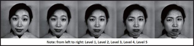
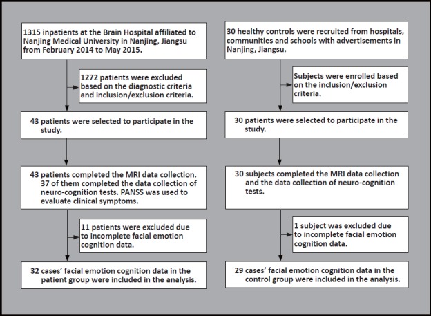
Methods: The neuro-cognitive functions of thirty-seven patients with first-episode schizophrenia (the patient group) and thirty healthy controls (the control group) was evaluated with the Clock Drawing Test, Trail Marking Test, Digit Span Test, Auditory Verbal Learning Test, Wisconsin Card Sorting Test, Verbal Fluency Test, Semantic Similarity Test and Stroop Color-Word Test. The facial emotion cognitive task was employed to assess the facial emotion cognitive functions of thirty-two patients with first-episode schizophrenia (the patient group) and 29 healthy controls (the control group). The psychotic symptoms of patients with first-episode schizophrenia were evaluated using the Positive and Negative Syndrome Scale (PANSS). The brain imaging data of the patient group and control group were collected using the magnetic resonance imagine (MRI).
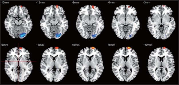
Results: The difference between the patient group and the control group in the results of Clock Drawing Test, Trail Marking Test, Digit Span Test, Auditory Verbal Learning Test, Wisconsin Card Sorting Test, Verbal Fluency Test, Semantic Similarity Test and Stroop Color-Word Test's reaction time were significant. These two groups' Slopes in the facial emotion cognitive task were also significantly different from each other. According to the comparison of cerebral grey matter volume between the patient group and the control group, it was found that the grey matter volume of the patient group increased in the left superior frontal gyrus, and decreased in the left occipital gyrus, lingual gyrus and upper cerebellum. Based on the analyses of neuro-cognitive data and brain imaging data of the patient group, the scores of the number of correct responses in Stroop Color-Word Test's Card C were negatively correlated with grey matter volumes of the left upper frontal gyrus, right upper frontal gyrus and middle frontal gyrus. The analyses on the facial emotion cognitive task and brain imaging data of the patient group showed that the slope data were positively correlated with grey matter volumes of the right superior temporal gyrus, middle temporal gyrus, left middle temporal gyrus, inferior temporal gyrus and fusiform gyrus.
Conclusion: There are general impairments in the neuro-cognitive functions and facial emotion cognitive functions of patients with first-episode schizophrenia, and the results suggest that brain areas with abnormal grey matter volumes are likely to be the brain structure and functional basis of the cognitive impairments.
期刊介绍:
Shanghai archives of psychiatry (bimonthly) was founded in 1959 and is sponsored by Shanghai Mental Health Center. The journal is aimed at mental health workers across the country, including psychiatrists and nurses, clinical psychologists, social workers, and people who are committed to the cause of mental health. It focuses on reporting clinical research results and practical experience in the field of psychiatry, and introduces the latest knowledge in psychiatry and related fields. The columns include monographs, case reports, clinical case discussions, reviews, mental health and law, and debates and discussions.


 分享
分享
 求助内容:
求助内容: 应助结果提醒方式:
 扫码关注我们
