{"title":"Insufficiency of collagenases in establishment of primary chondrocyte culture from cartilage of elderly patients receiving total joint replacement.","authors":"Jiamin Mao, Lexi Huang, Yiyang Ding, Xiaoyu Ma, Quanming Wang, Lei Ding","doi":"10.1007/s10561-023-10094-0","DOIUrl":null,"url":null,"abstract":"<p><p>Background Collagenases are frequently used in chondrocyte isolation from articular cartilage. However, the sufficiency of this enzyme in establishing primary human chondrocyte culture remains unknown. Methods Cartilage slices shaved from femoral head or tibial plateau of patients receiving total joint replacement surgery (16 hips, 8 knees) were subjected to 0.02% collagenase IA digestion for 16 h with (N = 19) or without (N = 5) the pre-treatment of 0.4% pronase E for 1.5 h. Chondrocyte yield and viability were compared between two groups. Chondrocyte phenotype was determined by the expression ratio of collagen type II to I. The morphology of cultured chondrocytes was monitored with a light microscope.Results Cartilage with pronase E pre-treatment yielded significantly higher chondrocytes than that without the pre-treatment (3,399 ± 1,637 cells/mg wet cartilage vs. 1,895 ± 688 cells/mg wet cartilage; P = 0.0067). Cell viability in the former group was also significantly higher than that in the latter (94% ± 2% vs. 86% ± 6%; P = 0.03). When cultured in monolayers, cells from cartilage with pronase E pre-treatment grew in a single plane showing rounded shape while cells from the other group grew in multi-planes and exhibited irregular shape. The mRNA expression ratio of collagen type II to I was 13.2 ± 7.5 in cells isolated from cartilage pre-treated with pronase E, indicating a typical chondrocyte phenotype. Conclusions Collagenase IA was not sufficient in establishing primary human chondrocyte culture. Cartilage must be treated with pronase E prior to collagenase IA application.</p>","PeriodicalId":9723,"journal":{"name":"Cell and Tissue Banking","volume":" ","pages":"759-768"},"PeriodicalIF":2.0000,"publicationDate":"2023-12-01","publicationTypes":"Journal Article","fieldsOfStudy":null,"isOpenAccess":false,"openAccessPdf":"","citationCount":"0","resultStr":null,"platform":"Semanticscholar","paperid":null,"PeriodicalName":"Cell and Tissue Banking","FirstCategoryId":"5","ListUrlMain":"https://doi.org/10.1007/s10561-023-10094-0","RegionNum":4,"RegionCategory":"医学","ArticlePicture":[],"TitleCN":null,"AbstractTextCN":null,"PMCID":null,"EPubDate":"2023/5/3 0:00:00","PubModel":"Epub","JCR":"Q4","JCRName":"CELL BIOLOGY","Score":null,"Total":0}
引用次数: 0
Abstract
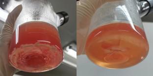
Background Collagenases are frequently used in chondrocyte isolation from articular cartilage. However, the sufficiency of this enzyme in establishing primary human chondrocyte culture remains unknown. Methods Cartilage slices shaved from femoral head or tibial plateau of patients receiving total joint replacement surgery (16 hips, 8 knees) were subjected to 0.02% collagenase IA digestion for 16 h with (N = 19) or without (N = 5) the pre-treatment of 0.4% pronase E for 1.5 h. Chondrocyte yield and viability were compared between two groups. Chondrocyte phenotype was determined by the expression ratio of collagen type II to I. The morphology of cultured chondrocytes was monitored with a light microscope.Results Cartilage with pronase E pre-treatment yielded significantly higher chondrocytes than that without the pre-treatment (3,399 ± 1,637 cells/mg wet cartilage vs. 1,895 ± 688 cells/mg wet cartilage; P = 0.0067). Cell viability in the former group was also significantly higher than that in the latter (94% ± 2% vs. 86% ± 6%; P = 0.03). When cultured in monolayers, cells from cartilage with pronase E pre-treatment grew in a single plane showing rounded shape while cells from the other group grew in multi-planes and exhibited irregular shape. The mRNA expression ratio of collagen type II to I was 13.2 ± 7.5 in cells isolated from cartilage pre-treated with pronase E, indicating a typical chondrocyte phenotype. Conclusions Collagenase IA was not sufficient in establishing primary human chondrocyte culture. Cartilage must be treated with pronase E prior to collagenase IA application.
期刊介绍:
Cell and Tissue Banking provides a forum for disseminating information to scientists and clinicians involved in the banking and transplantation of cells and tissues. Cell and Tissue Banking is an international, peer-reviewed journal that publishes original papers in the following areas:
basic research concerning general aspects of tissue banking such as quality assurance and control of banked cells/tissues, effects of preservation and sterilisation methods on cells/tissues, biotechnology, etc.; clinical applications of banked cells/tissues; standards of practice in procurement, processing, storage and distribution of cells/tissues; ethical issues; medico-legal issues.


 分享
分享
 求助内容:
求助内容: 应助结果提醒方式:
 扫码关注我们
