Alexander Pattyn , Yan Yan , Mohammad Mehrmohammadi
{"title":"环形阵列系统定量光谱光声层析成像的波长相关误差最小化。","authors":"Alexander Pattyn , Yan Yan , Mohammad Mehrmohammadi","doi":"10.1016/j.zemedi.2023.04.005","DOIUrl":null,"url":null,"abstract":"<div><h3>Purpose</h3><p>Photoacoustic tomography (PAT) is a non-invasive and high-resolution imaging technique that can provide functional and molecular information from the optical properties of pathological tissues, such as cancer. Spectroscopic PAT (sPAT) is capable of supplying information such as oxygen saturation (sO<sub>2</sub>), which is an important biological indicator for diseases such as cancer. However, the wavelength dependent nature of sPAT makes it challenging to provide accurate quantitative measurements of tissue oxygenation beyond shallow depths. We have previously reported the utility of combined ultrasound tomography and PAT to achieve optical and acoustic compensated PAT images at a single wavelength and for enhanced PAT images at larger depths. In this work we further explore the utility of the optical and acoustic compensation PAT algorithm to minimize the wavelength dependency in sPAT by showcasing improvements in spectral unmixing.</p></div><div><h3>Materials and Methods</h3><p>Two optically and acoustically characterized heterogenous phantoms were manufactured to test the ability of the system and developed algorithm to minimize the wavelength-dependence driven error in sPAT spectral unmixing. The PA inclusions within each phantom were composed of a mixture of two sulfate dyes, copper sulfate (CuSO<sub>4</sub>) and nickel sulfate (NiSO<sub>4</sub>), with known optical spectra. Improvements between uncompensated and optically and acoustically compensated PAT (OAcPAT) were quantified as the relative percent error between the measured results and the ground truth.</p></div><div><h3>Results</h3><p>The results of our phantom studies demonstrate that OAcPAT can significantly improve the accuracy of sPAT measurements in a heterogenous medium and especially at larger inclusions depths which can reach to up to 12% improvement in measurement errors. This significant improvement can play a vital role in reliability of future <em>in-vivo</em> biomarker quantifications.</p></div><div><h3>Conclusions</h3><p>Utilizing UST for model-based optical and acoustic compensation of PAT images was proposed by our group previously. In this work, we further demonstrated the efficacy of the developed algorithm in sPAT by minimizing the error caused by the tissue’s optical heterogeneity on improving spectral unmixing, which is a major limiting factor in reliability of sPAT measurements. Such synergistic combination of UST and PAT provides a window of opportunity to achieve bias-free quantitative sPAT measurements, which plays an important role in future pre-clinical and clinical utility of PAT.</p></div>","PeriodicalId":54397,"journal":{"name":"Zeitschrift fur Medizinische Physik","volume":"33 3","pages":"Pages 444-451"},"PeriodicalIF":4.2000,"publicationDate":"2023-08-01","publicationTypes":"Journal Article","fieldsOfStudy":null,"isOpenAccess":false,"openAccessPdf":"https://ftp.ncbi.nlm.nih.gov/pub/pmc/oa_pdf/6c/62/main.PMC10517392.pdf","citationCount":"0","resultStr":"{\"title\":\"Wavelength-dependent error minimization for quantitative spectroscopic photoacoustic tomography with a ring-array system\",\"authors\":\"Alexander Pattyn , Yan Yan , Mohammad Mehrmohammadi\",\"doi\":\"10.1016/j.zemedi.2023.04.005\",\"DOIUrl\":null,\"url\":null,\"abstract\":\"<div><h3>Purpose</h3><p>Photoacoustic tomography (PAT) is a non-invasive and high-resolution imaging technique that can provide functional and molecular information from the optical properties of pathological tissues, such as cancer. Spectroscopic PAT (sPAT) is capable of supplying information such as oxygen saturation (sO<sub>2</sub>), which is an important biological indicator for diseases such as cancer. However, the wavelength dependent nature of sPAT makes it challenging to provide accurate quantitative measurements of tissue oxygenation beyond shallow depths. We have previously reported the utility of combined ultrasound tomography and PAT to achieve optical and acoustic compensated PAT images at a single wavelength and for enhanced PAT images at larger depths. In this work we further explore the utility of the optical and acoustic compensation PAT algorithm to minimize the wavelength dependency in sPAT by showcasing improvements in spectral unmixing.</p></div><div><h3>Materials and Methods</h3><p>Two optically and acoustically characterized heterogenous phantoms were manufactured to test the ability of the system and developed algorithm to minimize the wavelength-dependence driven error in sPAT spectral unmixing. The PA inclusions within each phantom were composed of a mixture of two sulfate dyes, copper sulfate (CuSO<sub>4</sub>) and nickel sulfate (NiSO<sub>4</sub>), with known optical spectra. Improvements between uncompensated and optically and acoustically compensated PAT (OAcPAT) were quantified as the relative percent error between the measured results and the ground truth.</p></div><div><h3>Results</h3><p>The results of our phantom studies demonstrate that OAcPAT can significantly improve the accuracy of sPAT measurements in a heterogenous medium and especially at larger inclusions depths which can reach to up to 12% improvement in measurement errors. This significant improvement can play a vital role in reliability of future <em>in-vivo</em> biomarker quantifications.</p></div><div><h3>Conclusions</h3><p>Utilizing UST for model-based optical and acoustic compensation of PAT images was proposed by our group previously. In this work, we further demonstrated the efficacy of the developed algorithm in sPAT by minimizing the error caused by the tissue’s optical heterogeneity on improving spectral unmixing, which is a major limiting factor in reliability of sPAT measurements. Such synergistic combination of UST and PAT provides a window of opportunity to achieve bias-free quantitative sPAT measurements, which plays an important role in future pre-clinical and clinical utility of PAT.</p></div>\",\"PeriodicalId\":54397,\"journal\":{\"name\":\"Zeitschrift fur Medizinische Physik\",\"volume\":\"33 3\",\"pages\":\"Pages 444-451\"},\"PeriodicalIF\":4.2000,\"publicationDate\":\"2023-08-01\",\"publicationTypes\":\"Journal Article\",\"fieldsOfStudy\":null,\"isOpenAccess\":false,\"openAccessPdf\":\"https://ftp.ncbi.nlm.nih.gov/pub/pmc/oa_pdf/6c/62/main.PMC10517392.pdf\",\"citationCount\":\"0\",\"resultStr\":null,\"platform\":\"Semanticscholar\",\"paperid\":null,\"PeriodicalName\":\"Zeitschrift fur Medizinische Physik\",\"FirstCategoryId\":\"3\",\"ListUrlMain\":\"https://www.sciencedirect.com/science/article/pii/S0939388923000478\",\"RegionNum\":4,\"RegionCategory\":\"医学\",\"ArticlePicture\":[],\"TitleCN\":null,\"AbstractTextCN\":null,\"PMCID\":null,\"EPubDate\":\"2023/5/22 0:00:00\",\"PubModel\":\"Epub\",\"JCR\":\"Q2\",\"JCRName\":\"RADIOLOGY, NUCLEAR MEDICINE & MEDICAL IMAGING\",\"Score\":null,\"Total\":0}","platform":"Semanticscholar","paperid":null,"PeriodicalName":"Zeitschrift fur Medizinische Physik","FirstCategoryId":"3","ListUrlMain":"https://www.sciencedirect.com/science/article/pii/S0939388923000478","RegionNum":4,"RegionCategory":"医学","ArticlePicture":[],"TitleCN":null,"AbstractTextCN":null,"PMCID":null,"EPubDate":"2023/5/22 0:00:00","PubModel":"Epub","JCR":"Q2","JCRName":"RADIOLOGY, NUCLEAR MEDICINE & MEDICAL IMAGING","Score":null,"Total":0}
Wavelength-dependent error minimization for quantitative spectroscopic photoacoustic tomography with a ring-array system
Purpose
Photoacoustic tomography (PAT) is a non-invasive and high-resolution imaging technique that can provide functional and molecular information from the optical properties of pathological tissues, such as cancer. Spectroscopic PAT (sPAT) is capable of supplying information such as oxygen saturation (sO2), which is an important biological indicator for diseases such as cancer. However, the wavelength dependent nature of sPAT makes it challenging to provide accurate quantitative measurements of tissue oxygenation beyond shallow depths. We have previously reported the utility of combined ultrasound tomography and PAT to achieve optical and acoustic compensated PAT images at a single wavelength and for enhanced PAT images at larger depths. In this work we further explore the utility of the optical and acoustic compensation PAT algorithm to minimize the wavelength dependency in sPAT by showcasing improvements in spectral unmixing.
Materials and Methods
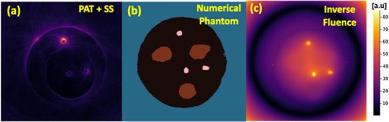
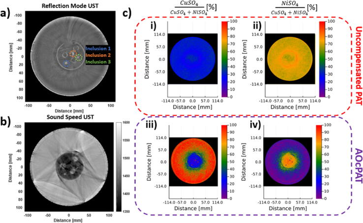
Two optically and acoustically characterized heterogenous phantoms were manufactured to test the ability of the system and developed algorithm to minimize the wavelength-dependence driven error in sPAT spectral unmixing. The PA inclusions within each phantom were composed of a mixture of two sulfate dyes, copper sulfate (CuSO4) and nickel sulfate (NiSO4), with known optical spectra. Improvements between uncompensated and optically and acoustically compensated PAT (OAcPAT) were quantified as the relative percent error between the measured results and the ground truth.
Results
The results of our phantom studies demonstrate that OAcPAT can significantly improve the accuracy of sPAT measurements in a heterogenous medium and especially at larger inclusions depths which can reach to up to 12% improvement in measurement errors. This significant improvement can play a vital role in reliability of future in-vivo biomarker quantifications.
Conclusions
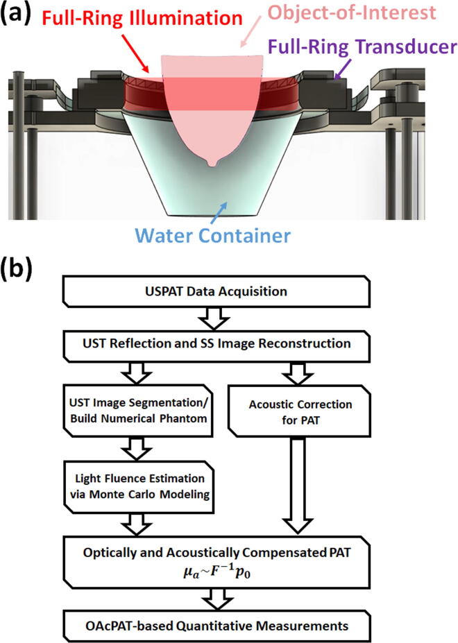
Utilizing UST for model-based optical and acoustic compensation of PAT images was proposed by our group previously. In this work, we further demonstrated the efficacy of the developed algorithm in sPAT by minimizing the error caused by the tissue’s optical heterogeneity on improving spectral unmixing, which is a major limiting factor in reliability of sPAT measurements. Such synergistic combination of UST and PAT provides a window of opportunity to achieve bias-free quantitative sPAT measurements, which plays an important role in future pre-clinical and clinical utility of PAT.
期刊介绍:
Zeitschrift fur Medizinische Physik (Journal of Medical Physics) is an official organ of the German and Austrian Society of Medical Physic and the Swiss Society of Radiobiology and Medical Physics.The Journal is a platform for basic research and practical applications of physical procedures in medical diagnostics and therapy. The articles are reviewed following international standards of peer reviewing.
Focuses of the articles are:
-Biophysical methods in radiation therapy and nuclear medicine
-Dosimetry and radiation protection
-Radiological diagnostics and quality assurance
-Modern imaging techniques, such as computed tomography, magnetic resonance imaging, positron emission tomography
-Ultrasonography diagnostics, application of laser and UV rays
-Electronic processing of biosignals
-Artificial intelligence and machine learning in medical physics
In the Journal, the latest scientific insights find their expression in the form of original articles, reviews, technical communications, and information for the clinical practice.


 分享
分享
 求助内容:
求助内容: 应助结果提醒方式:
 扫码关注我们
