{"title":"超声定位显微镜。","authors":"Stefanie Dencks, Georg Schmitz","doi":"10.1016/j.zemedi.2023.02.004","DOIUrl":null,"url":null,"abstract":"<div><p>Ultrasound Localization Microscopy (ULM) is an emerging technique that provides impressive super-resolved images of microvasculature, i.e., images with much better resolution than the conventional diffraction-limited ultrasound techniques and is already taking its first steps from preclinical to clinical applications. In comparison to the established perfusion or flow measurement methods, namely contrast-enhanced ultrasound (CEUS) and Doppler techniques, ULM allows imaging and flow measurements even down to the capillary level. As ULM can be realized as a post-processing method, conventional ultrasound systems can be used for.</p><p>ULM relies on the localization of single microbubbles (MB) of commercial, clinically approved contrast agents. In general, these very small and strong scatterers with typical radii of 1-3 <em>µm</em> are imaged much larger in ultrasound images than they actually are due to the point spread function of the imaging system. However, by applying appropriate methods, these MBs can be localized with sub-pixel precision. Then, by tracking MBs over successive frames of image sequences, not only the morphology of vascular trees but also functional information such as flow velocities or directions can be obtained and visualized. In addition, quantitative parameters can be derived to describe pathological and physiological changes in the microvasculature.</p><p>In this review, the general concept of ULM and conditions for its applicability to microvessel imaging are explained. Based on this, various aspects of the different processing steps for a concrete implementation are discussed. The trade-off between complete reconstruction of the microvasculature and the necessary measurement time as well as the implementation in 3D are reviewed in more detail, as they are the focus of current research. Through an overview of potential or already realized preclinical and clinical applications – pathologic angiogenesis or degeneration of vessels, physiological angiogenesis, or the general understanding of organ or tissue function – the great potential of ULM is demonstrated.</p></div>","PeriodicalId":54397,"journal":{"name":"Zeitschrift fur Medizinische Physik","volume":"33 3","pages":"Pages 292-308"},"PeriodicalIF":4.7000,"publicationDate":"2023-08-01","publicationTypes":"Journal Article","fieldsOfStudy":null,"isOpenAccess":false,"openAccessPdf":"https://www.ncbi.nlm.nih.gov/pmc/articles/PMC10517400/pdf/","citationCount":"2","resultStr":"{\"title\":\"Ultrasound localization microscopy\",\"authors\":\"Stefanie Dencks, Georg Schmitz\",\"doi\":\"10.1016/j.zemedi.2023.02.004\",\"DOIUrl\":null,\"url\":null,\"abstract\":\"<div><p>Ultrasound Localization Microscopy (ULM) is an emerging technique that provides impressive super-resolved images of microvasculature, i.e., images with much better resolution than the conventional diffraction-limited ultrasound techniques and is already taking its first steps from preclinical to clinical applications. In comparison to the established perfusion or flow measurement methods, namely contrast-enhanced ultrasound (CEUS) and Doppler techniques, ULM allows imaging and flow measurements even down to the capillary level. As ULM can be realized as a post-processing method, conventional ultrasound systems can be used for.</p><p>ULM relies on the localization of single microbubbles (MB) of commercial, clinically approved contrast agents. In general, these very small and strong scatterers with typical radii of 1-3 <em>µm</em> are imaged much larger in ultrasound images than they actually are due to the point spread function of the imaging system. However, by applying appropriate methods, these MBs can be localized with sub-pixel precision. Then, by tracking MBs over successive frames of image sequences, not only the morphology of vascular trees but also functional information such as flow velocities or directions can be obtained and visualized. In addition, quantitative parameters can be derived to describe pathological and physiological changes in the microvasculature.</p><p>In this review, the general concept of ULM and conditions for its applicability to microvessel imaging are explained. Based on this, various aspects of the different processing steps for a concrete implementation are discussed. The trade-off between complete reconstruction of the microvasculature and the necessary measurement time as well as the implementation in 3D are reviewed in more detail, as they are the focus of current research. Through an overview of potential or already realized preclinical and clinical applications – pathologic angiogenesis or degeneration of vessels, physiological angiogenesis, or the general understanding of organ or tissue function – the great potential of ULM is demonstrated.</p></div>\",\"PeriodicalId\":54397,\"journal\":{\"name\":\"Zeitschrift fur Medizinische Physik\",\"volume\":\"33 3\",\"pages\":\"Pages 292-308\"},\"PeriodicalIF\":4.7000,\"publicationDate\":\"2023-08-01\",\"publicationTypes\":\"Journal Article\",\"fieldsOfStudy\":null,\"isOpenAccess\":false,\"openAccessPdf\":\"https://www.ncbi.nlm.nih.gov/pmc/articles/PMC10517400/pdf/\",\"citationCount\":\"2\",\"resultStr\":null,\"platform\":\"Semanticscholar\",\"paperid\":null,\"PeriodicalName\":\"Zeitschrift fur Medizinische Physik\",\"FirstCategoryId\":\"3\",\"ListUrlMain\":\"https://www.sciencedirect.com/science/article/pii/S0939388923000302\",\"RegionNum\":4,\"RegionCategory\":\"医学\",\"ArticlePicture\":[],\"TitleCN\":null,\"AbstractTextCN\":null,\"PMCID\":null,\"EPubDate\":\"2023/6/15 0:00:00\",\"PubModel\":\"Epub\",\"JCR\":\"Q2\",\"JCRName\":\"RADIOLOGY, NUCLEAR MEDICINE & MEDICAL IMAGING\",\"Score\":null,\"Total\":0}","platform":"Semanticscholar","paperid":null,"PeriodicalName":"Zeitschrift fur Medizinische Physik","FirstCategoryId":"3","ListUrlMain":"https://www.sciencedirect.com/science/article/pii/S0939388923000302","RegionNum":4,"RegionCategory":"医学","ArticlePicture":[],"TitleCN":null,"AbstractTextCN":null,"PMCID":null,"EPubDate":"2023/6/15 0:00:00","PubModel":"Epub","JCR":"Q2","JCRName":"RADIOLOGY, NUCLEAR MEDICINE & MEDICAL IMAGING","Score":null,"Total":0}
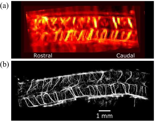
Ultrasound Localization Microscopy (ULM) is an emerging technique that provides impressive super-resolved images of microvasculature, i.e., images with much better resolution than the conventional diffraction-limited ultrasound techniques and is already taking its first steps from preclinical to clinical applications. In comparison to the established perfusion or flow measurement methods, namely contrast-enhanced ultrasound (CEUS) and Doppler techniques, ULM allows imaging and flow measurements even down to the capillary level. As ULM can be realized as a post-processing method, conventional ultrasound systems can be used for.
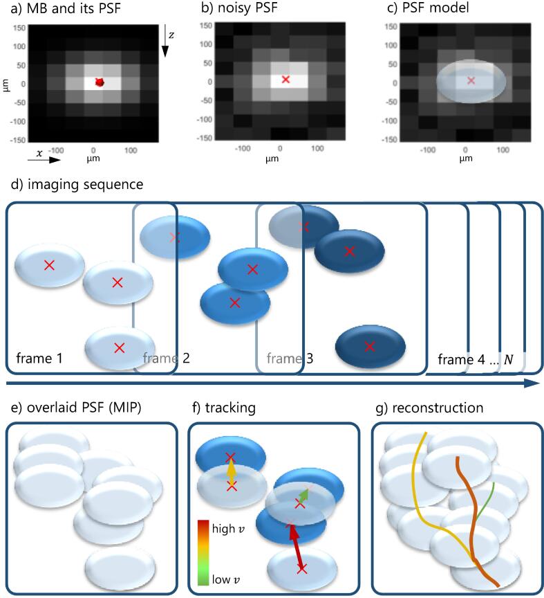
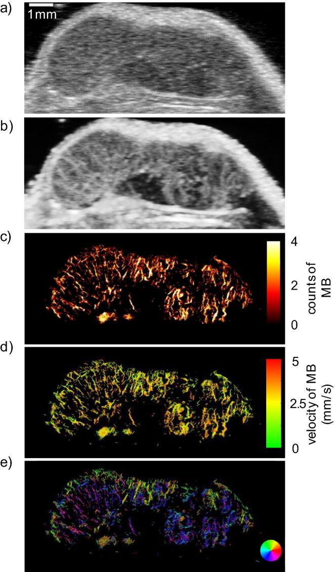
ULM relies on the localization of single microbubbles (MB) of commercial, clinically approved contrast agents. In general, these very small and strong scatterers with typical radii of 1-3 µm are imaged much larger in ultrasound images than they actually are due to the point spread function of the imaging system. However, by applying appropriate methods, these MBs can be localized with sub-pixel precision. Then, by tracking MBs over successive frames of image sequences, not only the morphology of vascular trees but also functional information such as flow velocities or directions can be obtained and visualized. In addition, quantitative parameters can be derived to describe pathological and physiological changes in the microvasculature.
In this review, the general concept of ULM and conditions for its applicability to microvessel imaging are explained. Based on this, various aspects of the different processing steps for a concrete implementation are discussed. The trade-off between complete reconstruction of the microvasculature and the necessary measurement time as well as the implementation in 3D are reviewed in more detail, as they are the focus of current research. Through an overview of potential or already realized preclinical and clinical applications – pathologic angiogenesis or degeneration of vessels, physiological angiogenesis, or the general understanding of organ or tissue function – the great potential of ULM is demonstrated.
期刊介绍:
Zeitschrift fur Medizinische Physik (Journal of Medical Physics) is an official organ of the German and Austrian Society of Medical Physic and the Swiss Society of Radiobiology and Medical Physics.The Journal is a platform for basic research and practical applications of physical procedures in medical diagnostics and therapy. The articles are reviewed following international standards of peer reviewing.
Focuses of the articles are:
-Biophysical methods in radiation therapy and nuclear medicine
-Dosimetry and radiation protection
-Radiological diagnostics and quality assurance
-Modern imaging techniques, such as computed tomography, magnetic resonance imaging, positron emission tomography
-Ultrasonography diagnostics, application of laser and UV rays
-Electronic processing of biosignals
-Artificial intelligence and machine learning in medical physics
In the Journal, the latest scientific insights find their expression in the form of original articles, reviews, technical communications, and information for the clinical practice.


 分享
分享
 求助内容:
求助内容: 应助结果提醒方式:
 扫码关注我们
