Eliška Materna Mikmeková , Jiří Materna , Ivo Konvalina , Šárka Mikmeková , Ilona Müllerová , Tewodros Asefa
{"title":"电子束的柔软触感:利用先进的扫描低能电子显微镜和深度学习挖掘纳米材料的结构信息","authors":"Eliška Materna Mikmeková , Jiří Materna , Ivo Konvalina , Šárka Mikmeková , Ilona Müllerová , Tewodros Asefa","doi":"10.1016/j.ultramic.2024.113965","DOIUrl":null,"url":null,"abstract":"<div><p>Nanostructured materials continue to find applications in various electronic and sensing devices, chromatography, separations, drug delivery, renewable energy, and catalysis. While major advancements on the synthesis and characterization of these materials have already been made, getting information about their structures at sub-nanometer resolution remains challenging. It is also unfortunate to find that many emerging or already available powerful analytical methods take time to be fully adopted for characterization of various nanomaterials. The scanning low energy electron microscopy (SLEEM) is a good example to this. In this report, we show how clearer structural and surface information at nanoscale can be obtained by SLEEM, coupled with deep learning. The method is demonstrated using Au nanoparticles-loaded mesoporous silica as a model system. Moreover, unlike conventional scanning electron microscopy (SEM), SLEEM does not require the samples to be coated with conductive films for analysis; thus, not only it is convenient to use but it also does not give artifacts. The results further reveal that SLEEM and deep learning can serve as great tools to analyze materials at nanoscale well. The biggest advantage of the presented method is its availability, as most modern SEMs are able to operate at low energies and deep learning methods are already being widely used in many fields.</p></div>","PeriodicalId":23439,"journal":{"name":"Ultramicroscopy","volume":"262 ","pages":"Article 113965"},"PeriodicalIF":2.0000,"publicationDate":"2024-08-01","publicationTypes":"Journal Article","fieldsOfStudy":null,"isOpenAccess":false,"openAccessPdf":"","citationCount":"0","resultStr":"{\"title\":\"A soft touch with electron beams: Digging out structural information of nanomaterials with advanced scanning low energy electron microscopy coupled with deep learning\",\"authors\":\"Eliška Materna Mikmeková , Jiří Materna , Ivo Konvalina , Šárka Mikmeková , Ilona Müllerová , Tewodros Asefa\",\"doi\":\"10.1016/j.ultramic.2024.113965\",\"DOIUrl\":null,\"url\":null,\"abstract\":\"<div><p>Nanostructured materials continue to find applications in various electronic and sensing devices, chromatography, separations, drug delivery, renewable energy, and catalysis. While major advancements on the synthesis and characterization of these materials have already been made, getting information about their structures at sub-nanometer resolution remains challenging. It is also unfortunate to find that many emerging or already available powerful analytical methods take time to be fully adopted for characterization of various nanomaterials. The scanning low energy electron microscopy (SLEEM) is a good example to this. In this report, we show how clearer structural and surface information at nanoscale can be obtained by SLEEM, coupled with deep learning. The method is demonstrated using Au nanoparticles-loaded mesoporous silica as a model system. Moreover, unlike conventional scanning electron microscopy (SEM), SLEEM does not require the samples to be coated with conductive films for analysis; thus, not only it is convenient to use but it also does not give artifacts. The results further reveal that SLEEM and deep learning can serve as great tools to analyze materials at nanoscale well. The biggest advantage of the presented method is its availability, as most modern SEMs are able to operate at low energies and deep learning methods are already being widely used in many fields.</p></div>\",\"PeriodicalId\":23439,\"journal\":{\"name\":\"Ultramicroscopy\",\"volume\":\"262 \",\"pages\":\"Article 113965\"},\"PeriodicalIF\":2.0000,\"publicationDate\":\"2024-08-01\",\"publicationTypes\":\"Journal Article\",\"fieldsOfStudy\":null,\"isOpenAccess\":false,\"openAccessPdf\":\"\",\"citationCount\":\"0\",\"resultStr\":null,\"platform\":\"Semanticscholar\",\"paperid\":null,\"PeriodicalName\":\"Ultramicroscopy\",\"FirstCategoryId\":\"5\",\"ListUrlMain\":\"https://www.sciencedirect.com/science/article/pii/S0304399124000445\",\"RegionNum\":3,\"RegionCategory\":\"工程技术\",\"ArticlePicture\":[],\"TitleCN\":null,\"AbstractTextCN\":null,\"PMCID\":null,\"EPubDate\":\"2024/4/10 0:00:00\",\"PubModel\":\"Epub\",\"JCR\":\"Q2\",\"JCRName\":\"MICROSCOPY\",\"Score\":null,\"Total\":0}","platform":"Semanticscholar","paperid":null,"PeriodicalName":"Ultramicroscopy","FirstCategoryId":"5","ListUrlMain":"https://www.sciencedirect.com/science/article/pii/S0304399124000445","RegionNum":3,"RegionCategory":"工程技术","ArticlePicture":[],"TitleCN":null,"AbstractTextCN":null,"PMCID":null,"EPubDate":"2024/4/10 0:00:00","PubModel":"Epub","JCR":"Q2","JCRName":"MICROSCOPY","Score":null,"Total":0}
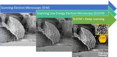
A soft touch with electron beams: Digging out structural information of nanomaterials with advanced scanning low energy electron microscopy coupled with deep learning
Nanostructured materials continue to find applications in various electronic and sensing devices, chromatography, separations, drug delivery, renewable energy, and catalysis. While major advancements on the synthesis and characterization of these materials have already been made, getting information about their structures at sub-nanometer resolution remains challenging. It is also unfortunate to find that many emerging or already available powerful analytical methods take time to be fully adopted for characterization of various nanomaterials. The scanning low energy electron microscopy (SLEEM) is a good example to this. In this report, we show how clearer structural and surface information at nanoscale can be obtained by SLEEM, coupled with deep learning. The method is demonstrated using Au nanoparticles-loaded mesoporous silica as a model system. Moreover, unlike conventional scanning electron microscopy (SEM), SLEEM does not require the samples to be coated with conductive films for analysis; thus, not only it is convenient to use but it also does not give artifacts. The results further reveal that SLEEM and deep learning can serve as great tools to analyze materials at nanoscale well. The biggest advantage of the presented method is its availability, as most modern SEMs are able to operate at low energies and deep learning methods are already being widely used in many fields.
期刊介绍:
Ultramicroscopy is an established journal that provides a forum for the publication of original research papers, invited reviews and rapid communications. The scope of Ultramicroscopy is to describe advances in instrumentation, methods and theory related to all modes of microscopical imaging, diffraction and spectroscopy in the life and physical sciences.


 分享
分享
 求助内容:
求助内容: 应助结果提醒方式:
 扫码关注我们
