Sejin Oh, Eunhye Yeo, Joonho Shim, Hyungrye Noh, Jihye Park, Kyeong-Tae Lee, Seok-Hyung Kim, Dongyoun Lee, Jong Hee Lee
{"title":"根据瘢痕疙瘩的状态揭示其发病机制:活跃与不活跃","authors":"Sejin Oh, Eunhye Yeo, Joonho Shim, Hyungrye Noh, Jihye Park, Kyeong-Tae Lee, Seok-Hyung Kim, Dongyoun Lee, Jong Hee Lee","doi":"10.1111/exd.15088","DOIUrl":null,"url":null,"abstract":"<p>Recently, the pathomechanisms of keloids have been extensively researched using transcriptomic analysis, but most studies did not consider the activity of keloids. We aimed to profile the transcriptomics of keloids according to their clinical activity and location within the keloid lesion, compared with normal and mature scars. Tissue samples were collected (keloid based on its activity (active and inactive), mature scar from keloid patients and normal scar (NS) from non-keloid patients). To reduce possible bias, all keloids assessed in this study had no treatment history and their location was limited to the upper chest or back. Multiomics assessment was performed by using single-cell RNA sequencing and multiplex immunofluorescence. Increased mesenchymal fibroblasts (FBs) was the main feature in keloid patients. Noticeably, the proportion of pro-inflammatory FBs was significantly increased in active keloids compared to inactive ones. To explore the nature of proinflammatory FBs, trajectory analysis was conducted and CCN family associated with mechanical stretch exhibited higher expression in active keloids. For vascular endothelial cells (VECs), the proportion of tip and immature cells increased in keloids compared to NS, especially at the periphery of active keloids. Also, keloid VECs highly expressed genes with characteristics of mesenchymal activation compared to NS, especially those from the active keloid center. Multiomics analysis demonstrated the distinct expression profile of active keloids. Clinically, these findings may provide the future appropriate directions for development of treatment modalities of keloids. Prevention of keloids could be possible by the suppression of mesenchymal activation between FBs and VECs and modulation of proinflammatory FBs may be the key to the control of active keloids.</p>","PeriodicalId":12243,"journal":{"name":"Experimental Dermatology","volume":"33 5","pages":""},"PeriodicalIF":3.1000,"publicationDate":"2024-04-29","publicationTypes":"Journal Article","fieldsOfStudy":null,"isOpenAccess":false,"openAccessPdf":"https://onlinelibrary.wiley.com/doi/epdf/10.1111/exd.15088","citationCount":"0","resultStr":"{\"title\":\"Revealing the pathogenesis of keloids based on the status: Active vs inactive\",\"authors\":\"Sejin Oh, Eunhye Yeo, Joonho Shim, Hyungrye Noh, Jihye Park, Kyeong-Tae Lee, Seok-Hyung Kim, Dongyoun Lee, Jong Hee Lee\",\"doi\":\"10.1111/exd.15088\",\"DOIUrl\":null,\"url\":null,\"abstract\":\"<p>Recently, the pathomechanisms of keloids have been extensively researched using transcriptomic analysis, but most studies did not consider the activity of keloids. We aimed to profile the transcriptomics of keloids according to their clinical activity and location within the keloid lesion, compared with normal and mature scars. Tissue samples were collected (keloid based on its activity (active and inactive), mature scar from keloid patients and normal scar (NS) from non-keloid patients). To reduce possible bias, all keloids assessed in this study had no treatment history and their location was limited to the upper chest or back. Multiomics assessment was performed by using single-cell RNA sequencing and multiplex immunofluorescence. Increased mesenchymal fibroblasts (FBs) was the main feature in keloid patients. Noticeably, the proportion of pro-inflammatory FBs was significantly increased in active keloids compared to inactive ones. To explore the nature of proinflammatory FBs, trajectory analysis was conducted and CCN family associated with mechanical stretch exhibited higher expression in active keloids. For vascular endothelial cells (VECs), the proportion of tip and immature cells increased in keloids compared to NS, especially at the periphery of active keloids. Also, keloid VECs highly expressed genes with characteristics of mesenchymal activation compared to NS, especially those from the active keloid center. Multiomics analysis demonstrated the distinct expression profile of active keloids. Clinically, these findings may provide the future appropriate directions for development of treatment modalities of keloids. Prevention of keloids could be possible by the suppression of mesenchymal activation between FBs and VECs and modulation of proinflammatory FBs may be the key to the control of active keloids.</p>\",\"PeriodicalId\":12243,\"journal\":{\"name\":\"Experimental Dermatology\",\"volume\":\"33 5\",\"pages\":\"\"},\"PeriodicalIF\":3.1000,\"publicationDate\":\"2024-04-29\",\"publicationTypes\":\"Journal Article\",\"fieldsOfStudy\":null,\"isOpenAccess\":false,\"openAccessPdf\":\"https://onlinelibrary.wiley.com/doi/epdf/10.1111/exd.15088\",\"citationCount\":\"0\",\"resultStr\":null,\"platform\":\"Semanticscholar\",\"paperid\":null,\"PeriodicalName\":\"Experimental Dermatology\",\"FirstCategoryId\":\"3\",\"ListUrlMain\":\"https://onlinelibrary.wiley.com/doi/10.1111/exd.15088\",\"RegionNum\":3,\"RegionCategory\":\"医学\",\"ArticlePicture\":[],\"TitleCN\":null,\"AbstractTextCN\":null,\"PMCID\":null,\"EPubDate\":\"\",\"PubModel\":\"\",\"JCR\":\"Q1\",\"JCRName\":\"DERMATOLOGY\",\"Score\":null,\"Total\":0}","platform":"Semanticscholar","paperid":null,"PeriodicalName":"Experimental Dermatology","FirstCategoryId":"3","ListUrlMain":"https://onlinelibrary.wiley.com/doi/10.1111/exd.15088","RegionNum":3,"RegionCategory":"医学","ArticlePicture":[],"TitleCN":null,"AbstractTextCN":null,"PMCID":null,"EPubDate":"","PubModel":"","JCR":"Q1","JCRName":"DERMATOLOGY","Score":null,"Total":0}
Revealing the pathogenesis of keloids based on the status: Active vs inactive
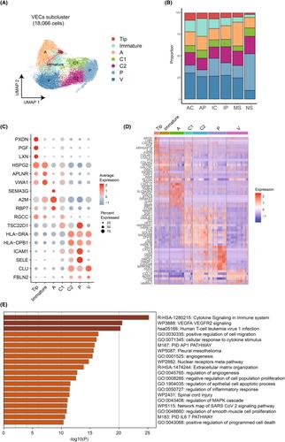
Recently, the pathomechanisms of keloids have been extensively researched using transcriptomic analysis, but most studies did not consider the activity of keloids. We aimed to profile the transcriptomics of keloids according to their clinical activity and location within the keloid lesion, compared with normal and mature scars. Tissue samples were collected (keloid based on its activity (active and inactive), mature scar from keloid patients and normal scar (NS) from non-keloid patients). To reduce possible bias, all keloids assessed in this study had no treatment history and their location was limited to the upper chest or back. Multiomics assessment was performed by using single-cell RNA sequencing and multiplex immunofluorescence. Increased mesenchymal fibroblasts (FBs) was the main feature in keloid patients. Noticeably, the proportion of pro-inflammatory FBs was significantly increased in active keloids compared to inactive ones. To explore the nature of proinflammatory FBs, trajectory analysis was conducted and CCN family associated with mechanical stretch exhibited higher expression in active keloids. For vascular endothelial cells (VECs), the proportion of tip and immature cells increased in keloids compared to NS, especially at the periphery of active keloids. Also, keloid VECs highly expressed genes with characteristics of mesenchymal activation compared to NS, especially those from the active keloid center. Multiomics analysis demonstrated the distinct expression profile of active keloids. Clinically, these findings may provide the future appropriate directions for development of treatment modalities of keloids. Prevention of keloids could be possible by the suppression of mesenchymal activation between FBs and VECs and modulation of proinflammatory FBs may be the key to the control of active keloids.
期刊介绍:
Experimental Dermatology provides a vehicle for the rapid publication of innovative and definitive reports, letters to the editor and review articles covering all aspects of experimental dermatology. Preference is given to papers of immediate importance to other investigators, either by virtue of their new methodology, experimental data or new ideas. The essential criteria for publication are clarity, experimental soundness and novelty. Letters to the editor related to published reports may also be accepted, provided that they are short and scientifically relevant to the reports mentioned, in order to provide a continuing forum for discussion. Review articles represent a state-of-the-art overview and are invited by the editors.


 分享
分享
 求助内容:
求助内容: 应助结果提醒方式:
 扫码关注我们
