{"title":"基于深度学习的 1.5 T 脑结构磁共振成像超分辨率:应用于定量体积测量。","authors":"Atita Suwannasak, Salita Angkurawaranon, Prapatsorn Sangpin, Itthi Chatnuntawech, Kittichai Wantanajittikul, Uten Yarach","doi":"10.1007/s10334-024-01165-8","DOIUrl":null,"url":null,"abstract":"<p><strong>Objective: </strong>This study investigated the feasibility of using deep learning-based super-resolution (DL-SR) technique on low-resolution (LR) images to generate high-resolution (HR) MR images with the aim of scan time reduction. The efficacy of DL-SR was also assessed through the application of brain volume measurement (BVM).</p><p><strong>Materials and methods: </strong>In vivo brain images acquired with 3D-T1W from various MRI scanners were utilized. For model training, LR images were generated by downsampling the original 1 mm-2 mm isotropic resolution images. Pairs of LR and HR images were used for training 3D residual dense net (RDN). For model testing, actual scanned 2 mm isotropic resolution 3D-T1W images with one-minute scan time were used. Normalized root-mean-square error (NRMSE), peak signal-to-noise ratio (PSNR), and structural similarity (SSIM) were used for model evaluation. The evaluation also included brain volume measurement, with assessments of subcortical brain regions.</p><p><strong>Results: </strong>The results showed that DL-SR model improved the quality of LR images compared with cubic interpolation, as indicated by NRMSE (24.22% vs 30.13%), PSNR (26.19 vs 24.65), and SSIM (0.96 vs 0.95). For volumetric assessments, there were no significant differences between DL-SR and actual HR images (p > 0.05, Pearson's correlation > 0.90) at seven subcortical regions.</p><p><strong>Discussion: </strong>The combination of LR MRI and DL-SR enables addressing prolonged scan time in 3D MRI scans while providing sufficient image quality without affecting brain volume measurement.</p>","PeriodicalId":18067,"journal":{"name":"Magnetic Resonance Materials in Physics, Biology and Medicine","volume":" ","pages":"465-475"},"PeriodicalIF":2.5000,"publicationDate":"2024-07-01","publicationTypes":"Journal Article","fieldsOfStudy":null,"isOpenAccess":false,"openAccessPdf":"","citationCount":"0","resultStr":"{\"title\":\"Deep learning-based super-resolution of structural brain MRI at 1.5 T: application to quantitative volume measurement.\",\"authors\":\"Atita Suwannasak, Salita Angkurawaranon, Prapatsorn Sangpin, Itthi Chatnuntawech, Kittichai Wantanajittikul, Uten Yarach\",\"doi\":\"10.1007/s10334-024-01165-8\",\"DOIUrl\":null,\"url\":null,\"abstract\":\"<p><strong>Objective: </strong>This study investigated the feasibility of using deep learning-based super-resolution (DL-SR) technique on low-resolution (LR) images to generate high-resolution (HR) MR images with the aim of scan time reduction. The efficacy of DL-SR was also assessed through the application of brain volume measurement (BVM).</p><p><strong>Materials and methods: </strong>In vivo brain images acquired with 3D-T1W from various MRI scanners were utilized. For model training, LR images were generated by downsampling the original 1 mm-2 mm isotropic resolution images. Pairs of LR and HR images were used for training 3D residual dense net (RDN). For model testing, actual scanned 2 mm isotropic resolution 3D-T1W images with one-minute scan time were used. Normalized root-mean-square error (NRMSE), peak signal-to-noise ratio (PSNR), and structural similarity (SSIM) were used for model evaluation. The evaluation also included brain volume measurement, with assessments of subcortical brain regions.</p><p><strong>Results: </strong>The results showed that DL-SR model improved the quality of LR images compared with cubic interpolation, as indicated by NRMSE (24.22% vs 30.13%), PSNR (26.19 vs 24.65), and SSIM (0.96 vs 0.95). For volumetric assessments, there were no significant differences between DL-SR and actual HR images (p > 0.05, Pearson's correlation > 0.90) at seven subcortical regions.</p><p><strong>Discussion: </strong>The combination of LR MRI and DL-SR enables addressing prolonged scan time in 3D MRI scans while providing sufficient image quality without affecting brain volume measurement.</p>\",\"PeriodicalId\":18067,\"journal\":{\"name\":\"Magnetic Resonance Materials in Physics, Biology and Medicine\",\"volume\":\" \",\"pages\":\"465-475\"},\"PeriodicalIF\":2.5000,\"publicationDate\":\"2024-07-01\",\"publicationTypes\":\"Journal Article\",\"fieldsOfStudy\":null,\"isOpenAccess\":false,\"openAccessPdf\":\"\",\"citationCount\":\"0\",\"resultStr\":null,\"platform\":\"Semanticscholar\",\"paperid\":null,\"PeriodicalName\":\"Magnetic Resonance Materials in Physics, Biology and Medicine\",\"FirstCategoryId\":\"3\",\"ListUrlMain\":\"https://doi.org/10.1007/s10334-024-01165-8\",\"RegionNum\":4,\"RegionCategory\":\"医学\",\"ArticlePicture\":[],\"TitleCN\":null,\"AbstractTextCN\":null,\"PMCID\":null,\"EPubDate\":\"2024/5/17 0:00:00\",\"PubModel\":\"Epub\",\"JCR\":\"Q3\",\"JCRName\":\"RADIOLOGY, NUCLEAR MEDICINE & MEDICAL IMAGING\",\"Score\":null,\"Total\":0}","platform":"Semanticscholar","paperid":null,"PeriodicalName":"Magnetic Resonance Materials in Physics, Biology and Medicine","FirstCategoryId":"3","ListUrlMain":"https://doi.org/10.1007/s10334-024-01165-8","RegionNum":4,"RegionCategory":"医学","ArticlePicture":[],"TitleCN":null,"AbstractTextCN":null,"PMCID":null,"EPubDate":"2024/5/17 0:00:00","PubModel":"Epub","JCR":"Q3","JCRName":"RADIOLOGY, NUCLEAR MEDICINE & MEDICAL IMAGING","Score":null,"Total":0}
Deep learning-based super-resolution of structural brain MRI at 1.5 T: application to quantitative volume measurement.
Objective: This study investigated the feasibility of using deep learning-based super-resolution (DL-SR) technique on low-resolution (LR) images to generate high-resolution (HR) MR images with the aim of scan time reduction. The efficacy of DL-SR was also assessed through the application of brain volume measurement (BVM).
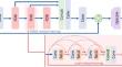
Materials and methods: In vivo brain images acquired with 3D-T1W from various MRI scanners were utilized. For model training, LR images were generated by downsampling the original 1 mm-2 mm isotropic resolution images. Pairs of LR and HR images were used for training 3D residual dense net (RDN). For model testing, actual scanned 2 mm isotropic resolution 3D-T1W images with one-minute scan time were used. Normalized root-mean-square error (NRMSE), peak signal-to-noise ratio (PSNR), and structural similarity (SSIM) were used for model evaluation. The evaluation also included brain volume measurement, with assessments of subcortical brain regions.
Results: The results showed that DL-SR model improved the quality of LR images compared with cubic interpolation, as indicated by NRMSE (24.22% vs 30.13%), PSNR (26.19 vs 24.65), and SSIM (0.96 vs 0.95). For volumetric assessments, there were no significant differences between DL-SR and actual HR images (p > 0.05, Pearson's correlation > 0.90) at seven subcortical regions.
Discussion: The combination of LR MRI and DL-SR enables addressing prolonged scan time in 3D MRI scans while providing sufficient image quality without affecting brain volume measurement.
期刊介绍:
MAGMA is a multidisciplinary international journal devoted to the publication of articles on all aspects of magnetic resonance techniques and their applications in medicine and biology. MAGMA currently publishes research papers, reviews, letters to the editor, and commentaries, six times a year. The subject areas covered by MAGMA include:
advances in materials, hardware and software in magnetic resonance technology,
new developments and results in research and practical applications of magnetic resonance imaging and spectroscopy related to biology and medicine,
study of animal models and intact cells using magnetic resonance,
reports of clinical trials on humans and clinical validation of magnetic resonance protocols.


 分享
分享

 求助内容:
求助内容: 应助结果提醒方式:
 扫码关注我们
