Man Wang , Muqi Jiang , Qi Wang , Yasheng Sun , Zhixiang Nie , William M. Palin , Zhen Zhang
{"title":"通过 i 型胶原蛋白的电泳沉积和仿生矿化提高钛植入物生物活性的体外生物启发方法。","authors":"Man Wang , Muqi Jiang , Qi Wang , Yasheng Sun , Zhixiang Nie , William M. Palin , Zhen Zhang","doi":"10.1016/j.bioadv.2024.214110","DOIUrl":null,"url":null,"abstract":"<div><h3>Objective</h3><div>This study aims to explore the efficacy of Electrophoretic Deposition (EPD) for collagen type I coating on titanium implants and its subsequent mineralization to improve osseointegration and bone regeneration.</div></div><div><h3>Methods</h3><div>Titanium disks were prepared with a sandblasted, large grit and acid-etched (SLA) surface. EPD was employed to deposit collagen type I onto the titanium surfaces, followed by two modes of mineralization: extra-fibril mineralization (EFM) and inter-fibril mineralization (IFM). Then comprehensive in vitro studies were conducted including surface properties, cell proliferation, osteogenic differentiation, and inflammatory responses.</div></div><div><h3>Results</h3><div>EPD successfully deposited a uniform collagen layer on titanium surfaces. EFM resulted in deposition of larger, irregularly shaped crystals, while IFM produced controlled, helical fibril mineralization. IFM-treated surfaces exhibited enhanced cell viability, proliferation, and osteogenic differentiation. Both EFM and IFM surfaces triggered higher macrophage activation than SLA surfaces. While EFM primarily induced a stronger M1 pro-inflammatory response, IFM exhibited a more balanced macrophage polarization with upregulated M2 markers at later stages.</div></div><div><h3>Conclusion</h3><div>EPD, particularly when integrated with IFM, significantly enhances the bioactivity and osteogenic potential of collagen-coated titanium implants. This method surpasses traditional SLA surfaces by stabilizing the collagen layer and creating a biomimetic environment conducive to bone regeneration and healing through a balanced inflammatory response, offering a promising strategy to improve titanium implant performance.</div></div>","PeriodicalId":51111,"journal":{"name":"Materials Science & Engineering C-Materials for Biological Applications","volume":"167 ","pages":"Article 214110"},"PeriodicalIF":6.0000,"publicationDate":"2025-02-01","publicationTypes":"Journal Article","fieldsOfStudy":null,"isOpenAccess":false,"openAccessPdf":"","citationCount":"0","resultStr":"{\"title\":\"An in vitro bioinspired approach to enhance the bioactivity of titanium implants via electrophoretic deposition and biomimetic mineralization of type i collagen\",\"authors\":\"Man Wang , Muqi Jiang , Qi Wang , Yasheng Sun , Zhixiang Nie , William M. Palin , Zhen Zhang\",\"doi\":\"10.1016/j.bioadv.2024.214110\",\"DOIUrl\":null,\"url\":null,\"abstract\":\"<div><h3>Objective</h3><div>This study aims to explore the efficacy of Electrophoretic Deposition (EPD) for collagen type I coating on titanium implants and its subsequent mineralization to improve osseointegration and bone regeneration.</div></div><div><h3>Methods</h3><div>Titanium disks were prepared with a sandblasted, large grit and acid-etched (SLA) surface. EPD was employed to deposit collagen type I onto the titanium surfaces, followed by two modes of mineralization: extra-fibril mineralization (EFM) and inter-fibril mineralization (IFM). Then comprehensive in vitro studies were conducted including surface properties, cell proliferation, osteogenic differentiation, and inflammatory responses.</div></div><div><h3>Results</h3><div>EPD successfully deposited a uniform collagen layer on titanium surfaces. EFM resulted in deposition of larger, irregularly shaped crystals, while IFM produced controlled, helical fibril mineralization. IFM-treated surfaces exhibited enhanced cell viability, proliferation, and osteogenic differentiation. Both EFM and IFM surfaces triggered higher macrophage activation than SLA surfaces. While EFM primarily induced a stronger M1 pro-inflammatory response, IFM exhibited a more balanced macrophage polarization with upregulated M2 markers at later stages.</div></div><div><h3>Conclusion</h3><div>EPD, particularly when integrated with IFM, significantly enhances the bioactivity and osteogenic potential of collagen-coated titanium implants. This method surpasses traditional SLA surfaces by stabilizing the collagen layer and creating a biomimetic environment conducive to bone regeneration and healing through a balanced inflammatory response, offering a promising strategy to improve titanium implant performance.</div></div>\",\"PeriodicalId\":51111,\"journal\":{\"name\":\"Materials Science & Engineering C-Materials for Biological Applications\",\"volume\":\"167 \",\"pages\":\"Article 214110\"},\"PeriodicalIF\":6.0000,\"publicationDate\":\"2025-02-01\",\"publicationTypes\":\"Journal Article\",\"fieldsOfStudy\":null,\"isOpenAccess\":false,\"openAccessPdf\":\"\",\"citationCount\":\"0\",\"resultStr\":null,\"platform\":\"Semanticscholar\",\"paperid\":null,\"PeriodicalName\":\"Materials Science & Engineering C-Materials for Biological Applications\",\"FirstCategoryId\":\"1085\",\"ListUrlMain\":\"https://www.sciencedirect.com/science/article/pii/S2772950824003534\",\"RegionNum\":2,\"RegionCategory\":\"医学\",\"ArticlePicture\":[],\"TitleCN\":null,\"AbstractTextCN\":null,\"PMCID\":null,\"EPubDate\":\"2024/11/8 0:00:00\",\"PubModel\":\"Epub\",\"JCR\":\"Q2\",\"JCRName\":\"MATERIALS SCIENCE, BIOMATERIALS\",\"Score\":null,\"Total\":0}","platform":"Semanticscholar","paperid":null,"PeriodicalName":"Materials Science & Engineering C-Materials for Biological Applications","FirstCategoryId":"1085","ListUrlMain":"https://www.sciencedirect.com/science/article/pii/S2772950824003534","RegionNum":2,"RegionCategory":"医学","ArticlePicture":[],"TitleCN":null,"AbstractTextCN":null,"PMCID":null,"EPubDate":"2024/11/8 0:00:00","PubModel":"Epub","JCR":"Q2","JCRName":"MATERIALS SCIENCE, BIOMATERIALS","Score":null,"Total":0}
引用次数: 0
摘要
研究目的本研究旨在探讨电泳沉积法(EPD)在钛植入物上涂覆 I 型胶原蛋白的效果,以及随后的矿化过程,以改善骨结合和骨再生:方法:制备的钛盘表面经过喷砂、大颗粒和酸蚀(SLA)处理。采用 EPD 将 I 型胶原蛋白沉积到钛表面,然后进行两种矿化模式:纤维外矿化(EFM)和纤维间矿化(IFM)。然后进行了全面的体外研究,包括表面特性、细胞增殖、成骨分化和炎症反应:结果:EPD 成功地在钛表面沉积了均匀的胶原层。EFM沉积了较大的不规则晶体,而IFM则产生了可控的螺旋状纤维矿化。经 IFM 处理的表面显示出更强的细胞活力、增殖和成骨分化能力。与 SLA 表面相比,EFM 和 IFM 表面都能引发更高的巨噬细胞活化。EFM主要诱导更强的M1促炎反应,而IFM则表现出更平衡的巨噬细胞极化,在后期阶段M2标记物上调:结论:EPD,尤其是与 IFM 结合使用时,可显著增强胶原涂层钛植入物的生物活性和成骨潜力。这种方法超越了传统的 SLA 表面,它稳定了胶原层,并通过平衡的炎症反应创造了有利于骨再生和愈合的仿生环境,为提高钛植入物的性能提供了一种前景广阔的策略。
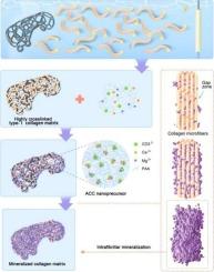
An in vitro bioinspired approach to enhance the bioactivity of titanium implants via electrophoretic deposition and biomimetic mineralization of type i collagen
Objective
This study aims to explore the efficacy of Electrophoretic Deposition (EPD) for collagen type I coating on titanium implants and its subsequent mineralization to improve osseointegration and bone regeneration.
Methods
Titanium disks were prepared with a sandblasted, large grit and acid-etched (SLA) surface. EPD was employed to deposit collagen type I onto the titanium surfaces, followed by two modes of mineralization: extra-fibril mineralization (EFM) and inter-fibril mineralization (IFM). Then comprehensive in vitro studies were conducted including surface properties, cell proliferation, osteogenic differentiation, and inflammatory responses.
Results
EPD successfully deposited a uniform collagen layer on titanium surfaces. EFM resulted in deposition of larger, irregularly shaped crystals, while IFM produced controlled, helical fibril mineralization. IFM-treated surfaces exhibited enhanced cell viability, proliferation, and osteogenic differentiation. Both EFM and IFM surfaces triggered higher macrophage activation than SLA surfaces. While EFM primarily induced a stronger M1 pro-inflammatory response, IFM exhibited a more balanced macrophage polarization with upregulated M2 markers at later stages.
Conclusion
EPD, particularly when integrated with IFM, significantly enhances the bioactivity and osteogenic potential of collagen-coated titanium implants. This method surpasses traditional SLA surfaces by stabilizing the collagen layer and creating a biomimetic environment conducive to bone regeneration and healing through a balanced inflammatory response, offering a promising strategy to improve titanium implant performance.
期刊介绍:
Biomaterials Advances, previously known as Materials Science and Engineering: C-Materials for Biological Applications (P-ISSN: 0928-4931, E-ISSN: 1873-0191). Includes topics at the interface of the biomedical sciences and materials engineering. These topics include:
• Bioinspired and biomimetic materials for medical applications
• Materials of biological origin for medical applications
• Materials for "active" medical applications
• Self-assembling and self-healing materials for medical applications
• "Smart" (i.e., stimulus-response) materials for medical applications
• Ceramic, metallic, polymeric, and composite materials for medical applications
• Materials for in vivo sensing
• Materials for in vivo imaging
• Materials for delivery of pharmacologic agents and vaccines
• Novel approaches for characterizing and modeling materials for medical applications
Manuscripts on biological topics without a materials science component, or manuscripts on materials science without biological applications, will not be considered for publication in Materials Science and Engineering C. New submissions are first assessed for language, scope and originality (plagiarism check) and can be desk rejected before review if they need English language improvements, are out of scope or present excessive duplication with published sources.
Biomaterials Advances sits within Elsevier''s biomaterials science portfolio alongside Biomaterials, Materials Today Bio and Biomaterials and Biosystems. As part of the broader Materials Today family, Biomaterials Advances offers authors rigorous peer review, rapid decisions, and high visibility. We look forward to receiving your submissions!


 分享
分享
 求助内容:
求助内容: 应助结果提醒方式:
 扫码关注我们
