Yodit Abebe Ayalew, Kinde Anlay Fante, Mohammed Aliy Mohammed
{"title":"用一种新的类平衡方法从计算机断层扫描图像中分割肝癌的改进型 U-Net","authors":"Yodit Abebe Ayalew, Kinde Anlay Fante, Mohammed Aliy Mohammed","doi":"10.1186/s42490-021-00050-y","DOIUrl":null,"url":null,"abstract":"<p><strong>Background: </strong>Liver cancer is the sixth most common cancer worldwide. It is mostly diagnosed with a computed tomography scan. Nowadays deep learning methods have been used for the segmentation of the liver and its tumor from the computed tomography (CT) scan images. This research mainly focused on segmenting liver and tumor from the abdominal CT scan images using a deep learning method and minimizing the effort and time used for a liver cancer diagnosis. The algorithm is based on the original UNet architecture. But, here in this paper, the numbers of filters on each convolutional block were reduced and new batch normalization and a dropout layer were added after each convolutional block of the contracting path.</p><p><strong>Results: </strong>Using this algorithm a dice score of 0.96, 0.74, and 0.63 were obtained for liver segmentation, segmentation of tumors from the liver, and the segmentation of tumor from abdominal CT scan images respectively. The segmentation results of liver and tumor from the liver showed an improvement of 0.01 and 0.11 respectively from other works.</p><p><strong>Conclusion: </strong>This work proposed a liver and a tumor segmentation method using a UNet architecture as a baseline. Modification regarding the number of filters and network layers were done on the original UNet model to reduce the network complexity and improve segmentation performance. A new class balancing method is also introduced to minimize the class imbalance problem. Through these, the algorithm attained better segmentation results and showed good improvement. However, it faced difficulty in segmenting small and irregular tumors.</p>","PeriodicalId":72425,"journal":{"name":"BMC biomedical engineering","volume":"3 1","pages":"4"},"PeriodicalIF":0.0000,"publicationDate":"2021-03-01","publicationTypes":"Journal Article","fieldsOfStudy":null,"isOpenAccess":false,"openAccessPdf":"https://www.ncbi.nlm.nih.gov/pmc/articles/PMC7919329/pdf/","citationCount":"0","resultStr":"{\"title\":\"Modified U-Net for liver cancer segmentation from computed tomography images with a new class balancing method.\",\"authors\":\"Yodit Abebe Ayalew, Kinde Anlay Fante, Mohammed Aliy Mohammed\",\"doi\":\"10.1186/s42490-021-00050-y\",\"DOIUrl\":null,\"url\":null,\"abstract\":\"<p><strong>Background: </strong>Liver cancer is the sixth most common cancer worldwide. It is mostly diagnosed with a computed tomography scan. Nowadays deep learning methods have been used for the segmentation of the liver and its tumor from the computed tomography (CT) scan images. This research mainly focused on segmenting liver and tumor from the abdominal CT scan images using a deep learning method and minimizing the effort and time used for a liver cancer diagnosis. The algorithm is based on the original UNet architecture. But, here in this paper, the numbers of filters on each convolutional block were reduced and new batch normalization and a dropout layer were added after each convolutional block of the contracting path.</p><p><strong>Results: </strong>Using this algorithm a dice score of 0.96, 0.74, and 0.63 were obtained for liver segmentation, segmentation of tumors from the liver, and the segmentation of tumor from abdominal CT scan images respectively. The segmentation results of liver and tumor from the liver showed an improvement of 0.01 and 0.11 respectively from other works.</p><p><strong>Conclusion: </strong>This work proposed a liver and a tumor segmentation method using a UNet architecture as a baseline. Modification regarding the number of filters and network layers were done on the original UNet model to reduce the network complexity and improve segmentation performance. A new class balancing method is also introduced to minimize the class imbalance problem. Through these, the algorithm attained better segmentation results and showed good improvement. However, it faced difficulty in segmenting small and irregular tumors.</p>\",\"PeriodicalId\":72425,\"journal\":{\"name\":\"BMC biomedical engineering\",\"volume\":\"3 1\",\"pages\":\"4\"},\"PeriodicalIF\":0.0000,\"publicationDate\":\"2021-03-01\",\"publicationTypes\":\"Journal Article\",\"fieldsOfStudy\":null,\"isOpenAccess\":false,\"openAccessPdf\":\"https://www.ncbi.nlm.nih.gov/pmc/articles/PMC7919329/pdf/\",\"citationCount\":\"0\",\"resultStr\":null,\"platform\":\"Semanticscholar\",\"paperid\":null,\"PeriodicalName\":\"BMC biomedical engineering\",\"FirstCategoryId\":\"1085\",\"ListUrlMain\":\"https://doi.org/10.1186/s42490-021-00050-y\",\"RegionNum\":0,\"RegionCategory\":null,\"ArticlePicture\":[],\"TitleCN\":null,\"AbstractTextCN\":null,\"PMCID\":null,\"EPubDate\":\"\",\"PubModel\":\"\",\"JCR\":\"\",\"JCRName\":\"\",\"Score\":null,\"Total\":0}","platform":"Semanticscholar","paperid":null,"PeriodicalName":"BMC biomedical engineering","FirstCategoryId":"1085","ListUrlMain":"https://doi.org/10.1186/s42490-021-00050-y","RegionNum":0,"RegionCategory":null,"ArticlePicture":[],"TitleCN":null,"AbstractTextCN":null,"PMCID":null,"EPubDate":"","PubModel":"","JCR":"","JCRName":"","Score":null,"Total":0}
Modified U-Net for liver cancer segmentation from computed tomography images with a new class balancing method.
Background: Liver cancer is the sixth most common cancer worldwide. It is mostly diagnosed with a computed tomography scan. Nowadays deep learning methods have been used for the segmentation of the liver and its tumor from the computed tomography (CT) scan images. This research mainly focused on segmenting liver and tumor from the abdominal CT scan images using a deep learning method and minimizing the effort and time used for a liver cancer diagnosis. The algorithm is based on the original UNet architecture. But, here in this paper, the numbers of filters on each convolutional block were reduced and new batch normalization and a dropout layer were added after each convolutional block of the contracting path.
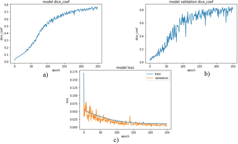
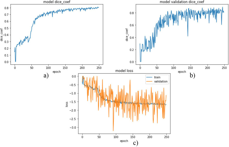
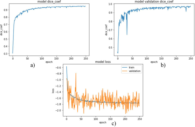
Results: Using this algorithm a dice score of 0.96, 0.74, and 0.63 were obtained for liver segmentation, segmentation of tumors from the liver, and the segmentation of tumor from abdominal CT scan images respectively. The segmentation results of liver and tumor from the liver showed an improvement of 0.01 and 0.11 respectively from other works.
Conclusion: This work proposed a liver and a tumor segmentation method using a UNet architecture as a baseline. Modification regarding the number of filters and network layers were done on the original UNet model to reduce the network complexity and improve segmentation performance. A new class balancing method is also introduced to minimize the class imbalance problem. Through these, the algorithm attained better segmentation results and showed good improvement. However, it faced difficulty in segmenting small and irregular tumors.


 分享
分享

 求助内容:
求助内容: 应助结果提醒方式:
 扫码关注我们
