Burak Oren, Gozde Aksoy Aydemir, Serkan Duzayak, Hasan Kızıltoprak
{"title":"光学相干断层扫描对斑秃视网膜层和脉络膜结构的评价。","authors":"Burak Oren, Gozde Aksoy Aydemir, Serkan Duzayak, Hasan Kızıltoprak","doi":"10.4274/MMJ.galenos.2023.58269","DOIUrl":null,"url":null,"abstract":"<p><strong>Objective: </strong>To evaluate the macula, retinal nerve fiber layer (RNFL), retinal layers, and choroidal thickness (CT) using spectral domain optical coherence tomography (SD-OCT) in patients with alopecia areata (AA).</p><p><strong>Methods: </strong>The right eyes of 42 AA patients (17 women, 25 men) and 42 controls (18 women, 24 men) were included in the study. Each subject underwent thorough ophthalmic examination and SD-OCT (Heidelberg Engineering) measurements. Central macular thickness (CMT), RNFL, the average thicknesses of the ganglion cell layer (GCL), inner plexiform layer (IPL), inner nuclear layer (INL), outer plexiform layer (OPL), outer nuclear layer (ONL), retinal pigment epithelium (RPE), inner retinal layers (IRL), photoreceptor layers (PRL) as well as subfoveal, temporal and nasal CT were measured.</p><p><strong>Results: </strong>In all sectors, no significant difference was observed between the AA group and the control group with regard to the mean values for CMT and RNFL (p>0.05, for all). There was not a significant difference between the AA group and the control group with regard to the thickness of the GCL, IPL, INL, OPL, ONL, RPE, IRL, and PRL (p>0.05 for all). CT at the subfoveal, temporal, and nasal regions was significantly thicker in the AA group than in the control group (p<0.05 for all).</p><p><strong>Conclusions: </strong>Along with T-lymphocyte-mediated hair follicle damage, choroidal melanocyte damage and inflammation can also be observed in AA patients. CT may increase secondary to melanocyte inflammation in AA patients.</p>","PeriodicalId":37427,"journal":{"name":"Medeniyet medical journal","volume":"38 2","pages":"140-147"},"PeriodicalIF":1.1000,"publicationDate":"2023-06-20","publicationTypes":"Journal Article","fieldsOfStudy":null,"isOpenAccess":false,"openAccessPdf":"https://ftp.ncbi.nlm.nih.gov/pub/pmc/oa_pdf/0f/ae/medj-38-140.PMC10284085.pdf","citationCount":"0","resultStr":"{\"title\":\"Evaluation of Retinal Layers and Choroidal Structures Using Optical Coherence Tomography in Alopecia Areata.\",\"authors\":\"Burak Oren, Gozde Aksoy Aydemir, Serkan Duzayak, Hasan Kızıltoprak\",\"doi\":\"10.4274/MMJ.galenos.2023.58269\",\"DOIUrl\":null,\"url\":null,\"abstract\":\"<p><strong>Objective: </strong>To evaluate the macula, retinal nerve fiber layer (RNFL), retinal layers, and choroidal thickness (CT) using spectral domain optical coherence tomography (SD-OCT) in patients with alopecia areata (AA).</p><p><strong>Methods: </strong>The right eyes of 42 AA patients (17 women, 25 men) and 42 controls (18 women, 24 men) were included in the study. Each subject underwent thorough ophthalmic examination and SD-OCT (Heidelberg Engineering) measurements. Central macular thickness (CMT), RNFL, the average thicknesses of the ganglion cell layer (GCL), inner plexiform layer (IPL), inner nuclear layer (INL), outer plexiform layer (OPL), outer nuclear layer (ONL), retinal pigment epithelium (RPE), inner retinal layers (IRL), photoreceptor layers (PRL) as well as subfoveal, temporal and nasal CT were measured.</p><p><strong>Results: </strong>In all sectors, no significant difference was observed between the AA group and the control group with regard to the mean values for CMT and RNFL (p>0.05, for all). There was not a significant difference between the AA group and the control group with regard to the thickness of the GCL, IPL, INL, OPL, ONL, RPE, IRL, and PRL (p>0.05 for all). CT at the subfoveal, temporal, and nasal regions was significantly thicker in the AA group than in the control group (p<0.05 for all).</p><p><strong>Conclusions: </strong>Along with T-lymphocyte-mediated hair follicle damage, choroidal melanocyte damage and inflammation can also be observed in AA patients. CT may increase secondary to melanocyte inflammation in AA patients.</p>\",\"PeriodicalId\":37427,\"journal\":{\"name\":\"Medeniyet medical journal\",\"volume\":\"38 2\",\"pages\":\"140-147\"},\"PeriodicalIF\":1.1000,\"publicationDate\":\"2023-06-20\",\"publicationTypes\":\"Journal Article\",\"fieldsOfStudy\":null,\"isOpenAccess\":false,\"openAccessPdf\":\"https://ftp.ncbi.nlm.nih.gov/pub/pmc/oa_pdf/0f/ae/medj-38-140.PMC10284085.pdf\",\"citationCount\":\"0\",\"resultStr\":null,\"platform\":\"Semanticscholar\",\"paperid\":null,\"PeriodicalName\":\"Medeniyet medical journal\",\"FirstCategoryId\":\"1085\",\"ListUrlMain\":\"https://doi.org/10.4274/MMJ.galenos.2023.58269\",\"RegionNum\":0,\"RegionCategory\":null,\"ArticlePicture\":[],\"TitleCN\":null,\"AbstractTextCN\":null,\"PMCID\":null,\"EPubDate\":\"\",\"PubModel\":\"\",\"JCR\":\"Q2\",\"JCRName\":\"MEDICINE, GENERAL & INTERNAL\",\"Score\":null,\"Total\":0}","platform":"Semanticscholar","paperid":null,"PeriodicalName":"Medeniyet medical journal","FirstCategoryId":"1085","ListUrlMain":"https://doi.org/10.4274/MMJ.galenos.2023.58269","RegionNum":0,"RegionCategory":null,"ArticlePicture":[],"TitleCN":null,"AbstractTextCN":null,"PMCID":null,"EPubDate":"","PubModel":"","JCR":"Q2","JCRName":"MEDICINE, GENERAL & INTERNAL","Score":null,"Total":0}
Evaluation of Retinal Layers and Choroidal Structures Using Optical Coherence Tomography in Alopecia Areata.
Objective: To evaluate the macula, retinal nerve fiber layer (RNFL), retinal layers, and choroidal thickness (CT) using spectral domain optical coherence tomography (SD-OCT) in patients with alopecia areata (AA).
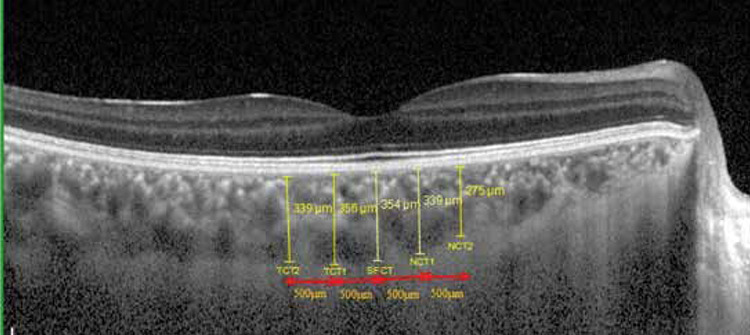
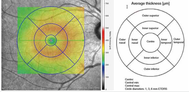
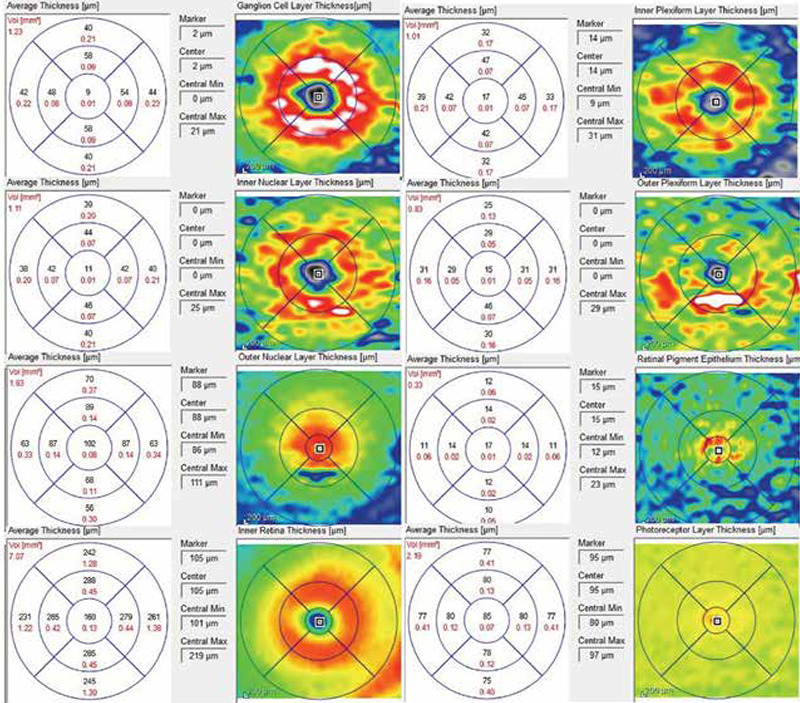
Methods: The right eyes of 42 AA patients (17 women, 25 men) and 42 controls (18 women, 24 men) were included in the study. Each subject underwent thorough ophthalmic examination and SD-OCT (Heidelberg Engineering) measurements. Central macular thickness (CMT), RNFL, the average thicknesses of the ganglion cell layer (GCL), inner plexiform layer (IPL), inner nuclear layer (INL), outer plexiform layer (OPL), outer nuclear layer (ONL), retinal pigment epithelium (RPE), inner retinal layers (IRL), photoreceptor layers (PRL) as well as subfoveal, temporal and nasal CT were measured.
Results: In all sectors, no significant difference was observed between the AA group and the control group with regard to the mean values for CMT and RNFL (p>0.05, for all). There was not a significant difference between the AA group and the control group with regard to the thickness of the GCL, IPL, INL, OPL, ONL, RPE, IRL, and PRL (p>0.05 for all). CT at the subfoveal, temporal, and nasal regions was significantly thicker in the AA group than in the control group (p<0.05 for all).
Conclusions: Along with T-lymphocyte-mediated hair follicle damage, choroidal melanocyte damage and inflammation can also be observed in AA patients. CT may increase secondary to melanocyte inflammation in AA patients.
期刊介绍:
The Medeniyet Medical Journal (Medeniyet Med J) is an open access, peer-reviewed, and scientific journal of Istanbul Medeniyet University Faculty of Medicine on various academic disciplines in medicine, which is published in English four times a year, in March, June, September, and December by a group of academics. Medeniyet Medical Journal is the continuation of Göztepe Medical Journal (ISSN: 1300-526X) which was started publishing in 1985. It changed the name as Medeniyet Medical Journal in 2015. Submission and publication are free of charge. No fees are asked from the authors for evaluation or publication process. All published articles are available online in the journal website (www.medeniyetmedicaljournal.org) without any fee. The journal publishes intradisciplinary or interdisciplinary clinical, experimental, and basic researches as well as original case reports, reviews, invited reviews, or letters to the editor, Being published since 1985, the Medeniyet Med J recognizes that the best science should lead to better lives based on the fact that the medicine should serve to the needs of society, and knowledge should transform society. The journal aims to address current issues at both national and international levels, start debates, and exert an influence on decision-makers all over the world by integrating science in everyday life. Medeniyet Med J is committed to serve the public and influence people’s lives in a positive way by making science widely accessible. Believing that the only goal is improving lives, and research has an impact on people’s lives, we select the best research papers in line with this goal.


 分享
分享
 求助内容:
求助内容: 应助结果提醒方式:
 扫码关注我们
