Burak Katipoglu, Nurullah İshak Işık, Ömer Faruk Turan, Safa Dönmez, Yusuf Yavuz, Ensar Durmuş, Attila Bestemir, Dariusz Timler
{"title":"A challenging decision for emergency physicians: Routine repeat computed brain tomography of the brain in head trauma in infants and neonates.","authors":"Burak Katipoglu, Nurullah İshak Işık, Ömer Faruk Turan, Safa Dönmez, Yusuf Yavuz, Ensar Durmuş, Attila Bestemir, Dariusz Timler","doi":"10.14744/tjtes.2024.28368","DOIUrl":null,"url":null,"abstract":"<p><strong>Background: </strong>Head trauma is a leading cause of death and disability. While standard treatment protocols exist for severe head trauma, no clear follow-up standards are available for mild head trauma with positive imaging findings in infants and newborns. Although routine follow-up brain computed tomography (CT) imaging is not recommended for children with moderate and mild head trauma, the necessity for follow-up imaging in infants and newborns remains uncertain.</p><p><strong>Methods: </strong>Our study is a retrospective, observational, and descriptive study. Infants under 1 year old presenting to the emergency department with isolated head trauma were reviewed with the approval of the Ethics Committee of Ankara Etlik City Hospital. Inclusion criteria included presentation to the emergency department, undergoing more than one brain CT scan, and sustaining mild head trauma (Glasgow Coma Scale [GCS] >13). Patients with incomplete follow-up data or multiple traumas were excluded. Age, gender, mechanism of trauma, initial and follow-up brain CT findings, hospital admission, and surgical procedures were recorded and analyzed using the SPSS statistical package.</p><p><strong>Results: </strong>Out of 238 screened patients, 154 were included in the study. Of these, 66.9% were male and the average age was 5.99 months. The most common presenting symptom was swelling at the trauma site, observed in 79.2% of cases. The most common mechanism of injury was falling from a height of less than 90 cm, accounting for 85.1% of cases. Pathological progression on follow-up CT was observed in 5.2% of the patients, and only 1.9% required surgical treatment. A total of 34.4% of the patients required hospitalization. Patients with parenchymal brain pathology had a higher rate of pathological progression on follow-up CT and a longer hospital stay.</p><p><strong>Conclusion: </strong>Follow-up CT scans in infants with mild head trauma do not alter patient outcomes except in cases with brain parenchymal pathology. Study data indicated that repeat imaging is not beneficial for isolated skull fractures. Imaging artifacts often necessitated repeated scans, contributing to increased radiation exposure. Unnecessary repeat imaging escalates radiation exposure and healthcare costs. Only a small percentage of patients exhibited progression of intracranial pathology, justifying follow-up imaging solely in the presence of brain parenchymal injury. Larger prospective studies are necessary to confirm these findings.</p>","PeriodicalId":94263,"journal":{"name":"Ulusal travma ve acil cerrahi dergisi = Turkish journal of trauma & emergency surgery : TJTES","volume":"30 8","pages":"596-602"},"PeriodicalIF":1.0000,"publicationDate":"2024-08-01","publicationTypes":"Journal Article","fieldsOfStudy":null,"isOpenAccess":false,"openAccessPdf":"https://www.ncbi.nlm.nih.gov/pmc/articles/PMC11372490/pdf/","citationCount":"0","resultStr":null,"platform":"Semanticscholar","paperid":null,"PeriodicalName":"Ulusal travma ve acil cerrahi dergisi = Turkish journal of trauma & emergency surgery : TJTES","FirstCategoryId":"1085","ListUrlMain":"https://doi.org/10.14744/tjtes.2024.28368","RegionNum":0,"RegionCategory":null,"ArticlePicture":[],"TitleCN":null,"AbstractTextCN":null,"PMCID":null,"EPubDate":"","PubModel":"","JCR":"","JCRName":"","Score":null,"Total":0}
引用次数: 0
Abstract
Background: Head trauma is a leading cause of death and disability. While standard treatment protocols exist for severe head trauma, no clear follow-up standards are available for mild head trauma with positive imaging findings in infants and newborns. Although routine follow-up brain computed tomography (CT) imaging is not recommended for children with moderate and mild head trauma, the necessity for follow-up imaging in infants and newborns remains uncertain.
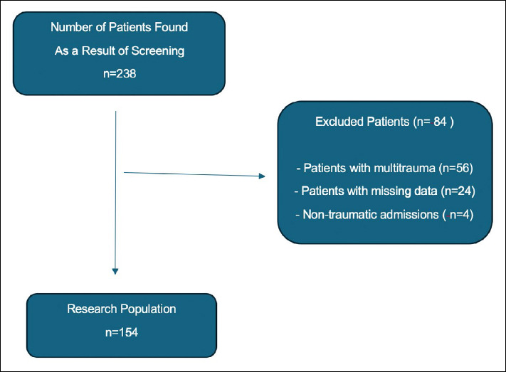
Methods: Our study is a retrospective, observational, and descriptive study. Infants under 1 year old presenting to the emergency department with isolated head trauma were reviewed with the approval of the Ethics Committee of Ankara Etlik City Hospital. Inclusion criteria included presentation to the emergency department, undergoing more than one brain CT scan, and sustaining mild head trauma (Glasgow Coma Scale [GCS] >13). Patients with incomplete follow-up data or multiple traumas were excluded. Age, gender, mechanism of trauma, initial and follow-up brain CT findings, hospital admission, and surgical procedures were recorded and analyzed using the SPSS statistical package.
Results: Out of 238 screened patients, 154 were included in the study. Of these, 66.9% were male and the average age was 5.99 months. The most common presenting symptom was swelling at the trauma site, observed in 79.2% of cases. The most common mechanism of injury was falling from a height of less than 90 cm, accounting for 85.1% of cases. Pathological progression on follow-up CT was observed in 5.2% of the patients, and only 1.9% required surgical treatment. A total of 34.4% of the patients required hospitalization. Patients with parenchymal brain pathology had a higher rate of pathological progression on follow-up CT and a longer hospital stay.
Conclusion: Follow-up CT scans in infants with mild head trauma do not alter patient outcomes except in cases with brain parenchymal pathology. Study data indicated that repeat imaging is not beneficial for isolated skull fractures. Imaging artifacts often necessitated repeated scans, contributing to increased radiation exposure. Unnecessary repeat imaging escalates radiation exposure and healthcare costs. Only a small percentage of patients exhibited progression of intracranial pathology, justifying follow-up imaging solely in the presence of brain parenchymal injury. Larger prospective studies are necessary to confirm these findings.


 分享
分享

 求助内容:
求助内容: 应助结果提醒方式:
 扫码关注我们
