{"title":"Liver fibrosis stage classification in stacked microvascular images based on deep learning.","authors":"Daisuke Miura, Hiromi Suenaga, Rino Hiwatashi, Shingo Mabu","doi":"10.1186/s12880-024-01531-x","DOIUrl":null,"url":null,"abstract":"<p><strong>Background: </strong>Monitoring fibrosis in patients with chronic liver disease (CLD) is an important management strategy. We have already reported a novel stacked microvascular imaging (SMVI) technique and an examiner scoring evaluation method to improve fibrosis assessment accuracy and demonstrate its high sensitivity. In the present study, we analyzed the effectiveness and objectivity of SMVI in diagnosing the liver fibrosis stage based on artificial intelligence (AI).</p><p><strong>Methods: </strong>This single-center, cross-sectional study included 517 patients with CLD who underwent ultrasonography and liver stiffness testing between August 2019 and October 2022. A convolutional neural network model was constructed to evaluate the degree of liver fibrosis from stacked microvascular images generated by accumulating high-sensitivity Doppler (i.e., high-definition color) images from these patients. In contrast, as a method of judgment by the human eye, we focused on three hallmarks of intrahepatic microvessel morphological changes in the stacked microvascular images: narrowing, caliber irregularity, and tortuosity. The degree of liver fibrosis was classified into five stages according to etiology based on liver stiffness measurement: F0-1Low (< 5.0 kPa), F0-1High (≥ 5.0 kPa), F2, F3, and F4.</p><p><strong>Results: </strong>The AI classification accuracy was 53.8% for a 5-class classification, 66.3% for a 3-class classification (F0-1Low vs. F0-1High vs. F2-4), and 83.8% for a 2-class classification (F0-1 vs. F2-4). The diagnostic accuracy for ≥ F2 was 81.6% in the examiner's score assessment, compared with 83.8% in AI assessment, indicating that AI achieved higher diagnostic accuracy. Similarly, AI demonstrated higher sensitivity and specificity of 84.2% and 83.5%, respectively. Comparing human judgement with AI judgement, the AI analysis was a superior model with a higher F1 score in the 2-class classification.</p><p><strong>Conclusions: </strong>In detecting significant fibrosis (≥ F2) using the SMVI method, AI-based assessments are more accurate than human judgement; moreover, AI-based SMVI analysis eliminating human subjectivity bias and determining patients with objective fibrosis development is considered an important improvement.</p>","PeriodicalId":9020,"journal":{"name":"BMC Medical Imaging","volume":"25 1","pages":"8"},"PeriodicalIF":3.2000,"publicationDate":"2025-01-07","publicationTypes":"Journal Article","fieldsOfStudy":null,"isOpenAccess":false,"openAccessPdf":"https://www.ncbi.nlm.nih.gov/pmc/articles/PMC11706143/pdf/","citationCount":"0","resultStr":null,"platform":"Semanticscholar","paperid":null,"PeriodicalName":"BMC Medical Imaging","FirstCategoryId":"3","ListUrlMain":"https://doi.org/10.1186/s12880-024-01531-x","RegionNum":3,"RegionCategory":"医学","ArticlePicture":[],"TitleCN":null,"AbstractTextCN":null,"PMCID":null,"EPubDate":"","PubModel":"","JCR":"Q2","JCRName":"RADIOLOGY, NUCLEAR MEDICINE & MEDICAL IMAGING","Score":null,"Total":0}
引用次数: 0
Abstract
Background: Monitoring fibrosis in patients with chronic liver disease (CLD) is an important management strategy. We have already reported a novel stacked microvascular imaging (SMVI) technique and an examiner scoring evaluation method to improve fibrosis assessment accuracy and demonstrate its high sensitivity. In the present study, we analyzed the effectiveness and objectivity of SMVI in diagnosing the liver fibrosis stage based on artificial intelligence (AI).
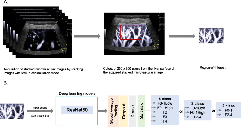
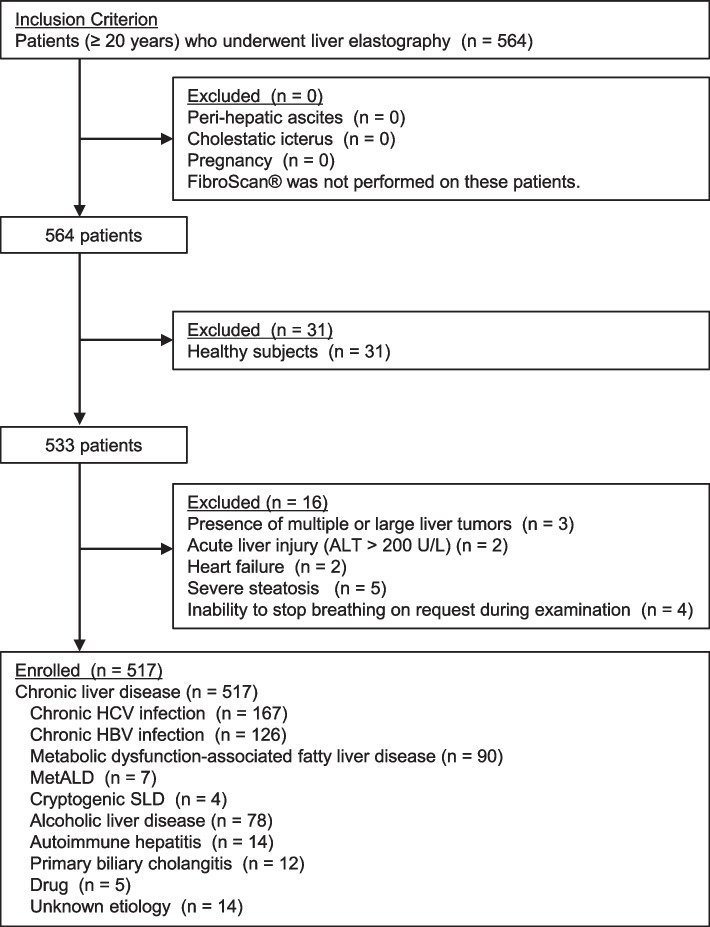
Methods: This single-center, cross-sectional study included 517 patients with CLD who underwent ultrasonography and liver stiffness testing between August 2019 and October 2022. A convolutional neural network model was constructed to evaluate the degree of liver fibrosis from stacked microvascular images generated by accumulating high-sensitivity Doppler (i.e., high-definition color) images from these patients. In contrast, as a method of judgment by the human eye, we focused on three hallmarks of intrahepatic microvessel morphological changes in the stacked microvascular images: narrowing, caliber irregularity, and tortuosity. The degree of liver fibrosis was classified into five stages according to etiology based on liver stiffness measurement: F0-1Low (< 5.0 kPa), F0-1High (≥ 5.0 kPa), F2, F3, and F4.
Results: The AI classification accuracy was 53.8% for a 5-class classification, 66.3% for a 3-class classification (F0-1Low vs. F0-1High vs. F2-4), and 83.8% for a 2-class classification (F0-1 vs. F2-4). The diagnostic accuracy for ≥ F2 was 81.6% in the examiner's score assessment, compared with 83.8% in AI assessment, indicating that AI achieved higher diagnostic accuracy. Similarly, AI demonstrated higher sensitivity and specificity of 84.2% and 83.5%, respectively. Comparing human judgement with AI judgement, the AI analysis was a superior model with a higher F1 score in the 2-class classification.
Conclusions: In detecting significant fibrosis (≥ F2) using the SMVI method, AI-based assessments are more accurate than human judgement; moreover, AI-based SMVI analysis eliminating human subjectivity bias and determining patients with objective fibrosis development is considered an important improvement.
背景:监测慢性肝病(CLD)患者的纤维化是一项重要的管理策略。我们已经报道了一种新的堆叠微血管成像(SMVI)技术和一种检查者评分评估方法,以提高纤维化评估的准确性并证明其高灵敏度。在本研究中,我们分析了基于人工智能(AI)的SMVI诊断肝纤维化分期的有效性和客观性。方法:这项单中心横断面研究包括517例CLD患者,他们在2019年8月至2022年10月期间接受了超声检查和肝脏硬度测试。构建卷积神经网络模型,通过累积这些患者的高灵敏度多普勒(即高清彩色)图像生成的堆叠微血管图像来评估肝纤维化程度。相比之下,作为人眼判断的方法,我们重点关注了堆积的微血管图像中肝内微血管形态变化的三个特征:狭窄、口径不规则和扭曲。基于肝硬度测量,根据病因将肝纤维化程度分为5个阶段:F0-1Low(结果:5级AI分类准确率为53.8%,3级AI分类准确率为66.3% (F0-1Low vs F0-1High vs F2-4), 2级AI分类准确率为83.8% (F0-1 vs F2-4)。在主考官评分评估中,≥F2的诊断准确率为81.6%,而人工智能评估的诊断准确率为83.8%,表明人工智能的诊断准确率更高。同样,AI具有更高的敏感性和特异性,分别为84.2%和83.5%。人工判断与人工智能判断相比,人工智能分析在2类分类中具有更高的F1分,是一种更优的模型。结论:在使用SMVI方法检测显著纤维化(≥F2)时,基于人工智能的评估比人类判断更准确;此外,基于人工智能的SMVI分析消除了人为的主观性偏差,并确定了客观纤维化发展的患者,被认为是一个重要的改进。
期刊介绍:
BMC Medical Imaging is an open access journal publishing original peer-reviewed research articles in the development, evaluation, and use of imaging techniques and image processing tools to diagnose and manage disease.


 分享
分享
 求助内容:
求助内容: 应助结果提醒方式:
 扫码关注我们
