Deep learning-based fully automated detection and segmentation of pelvic lymph nodes on diffusion-weighted images for prostate cancer: a multicenter study.
Zhaonan Sun, Pengsheng Wu, Tongtong Zhao, Ge Gao, Huihui Wang, Xiaodong Zhang, Xiaoying Wang
{"title":"Deep learning-based fully automated detection and segmentation of pelvic lymph nodes on diffusion-weighted images for prostate cancer: a multicenter study.","authors":"Zhaonan Sun, Pengsheng Wu, Tongtong Zhao, Ge Gao, Huihui Wang, Xiaodong Zhang, Xiaoying Wang","doi":"10.1186/s40644-025-00840-w","DOIUrl":null,"url":null,"abstract":"<p><strong>Background: </strong>Accurate identification and evaluation of lymph nodes (LNs) in prostate cancer (PCa) patients is crucial for effective staging but can be time-consuming. We utilized a 3D V-Net model to improve the efficiency and accuracy of LN detection and segmentation.</p><p><strong>Methods: </strong>Utilizing pelvic diffusion-weighted imaging (DWI) scans, the 3D V-Net framework underwent training on a dataset comprising data from a hospital with 1,151 patients, encompassing 32,507 annotated LNs, following data augmentation procedures. Subsequently, external validation was conducted on data from 401 patients across three additional hospitals, encompassing 7,707 LNs. The segmentation performance was evaluated using the Dice similarity coefficient (DSC). The comparison between automated and manual segmentation regarding the short diameter and volume of LNs was conducted using Bland-Altman plots and correlation analysis. The performance for suspicious metastatic LN detection (short diameter > 8 mm) was evaluated using sensitivity, positive predictive value (PPV), and per-patient false-positive rate (FP/vol) at the LN level and sensitivity, specificity, and PPV at the patient level.</p><p><strong>Results: </strong>In the external validation test dataset, the model achieved a DSC of 0.77-0.82 for all, suspicious, and largest LNs. The model achieved a sensitivity, PPV, and FP/vol of 60.1% (95% confidence interval (CI), 57.6-62.6%), 79.2% (95% CI, 76.6-81.5%), and 0.56 at the LN level, respectively. At the patient level, the model achieved a sensitivity, specificity, and PPV of 81.1% (95% CI, 76.5-85.0%), 75.6% (95% CI, 65.1-83.8%), and 93.2% (95% CI, 89.7-95.6%), respectively. The model achieved a strong correlation and good consistency between the short diameter and volume of the automatically segmented and manually annotated LNs.</p><p><strong>Conclusion: </strong>This 3D V-Net model can segment LNs effectively based on pelvic DWI images for PCa and holds great potential for facilitating N-staging in clinical practice.</p>","PeriodicalId":9548,"journal":{"name":"Cancer Imaging","volume":"25 1","pages":"37"},"PeriodicalIF":3.5000,"publicationDate":"2025-03-17","publicationTypes":"Journal Article","fieldsOfStudy":null,"isOpenAccess":false,"openAccessPdf":"https://www.ncbi.nlm.nih.gov/pmc/articles/PMC11912796/pdf/","citationCount":"0","resultStr":null,"platform":"Semanticscholar","paperid":null,"PeriodicalName":"Cancer Imaging","FirstCategoryId":"3","ListUrlMain":"https://doi.org/10.1186/s40644-025-00840-w","RegionNum":2,"RegionCategory":"医学","ArticlePicture":[],"TitleCN":null,"AbstractTextCN":null,"PMCID":null,"EPubDate":"","PubModel":"","JCR":"Q2","JCRName":"ONCOLOGY","Score":null,"Total":0}
引用次数: 0
Abstract
Background: Accurate identification and evaluation of lymph nodes (LNs) in prostate cancer (PCa) patients is crucial for effective staging but can be time-consuming. We utilized a 3D V-Net model to improve the efficiency and accuracy of LN detection and segmentation.
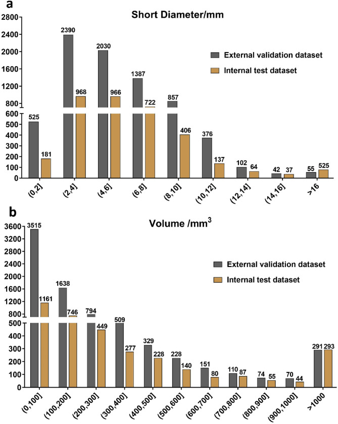
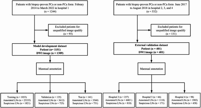
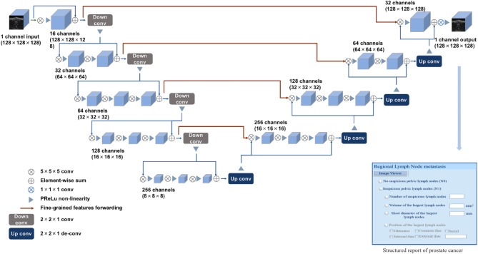
Methods: Utilizing pelvic diffusion-weighted imaging (DWI) scans, the 3D V-Net framework underwent training on a dataset comprising data from a hospital with 1,151 patients, encompassing 32,507 annotated LNs, following data augmentation procedures. Subsequently, external validation was conducted on data from 401 patients across three additional hospitals, encompassing 7,707 LNs. The segmentation performance was evaluated using the Dice similarity coefficient (DSC). The comparison between automated and manual segmentation regarding the short diameter and volume of LNs was conducted using Bland-Altman plots and correlation analysis. The performance for suspicious metastatic LN detection (short diameter > 8 mm) was evaluated using sensitivity, positive predictive value (PPV), and per-patient false-positive rate (FP/vol) at the LN level and sensitivity, specificity, and PPV at the patient level.
Results: In the external validation test dataset, the model achieved a DSC of 0.77-0.82 for all, suspicious, and largest LNs. The model achieved a sensitivity, PPV, and FP/vol of 60.1% (95% confidence interval (CI), 57.6-62.6%), 79.2% (95% CI, 76.6-81.5%), and 0.56 at the LN level, respectively. At the patient level, the model achieved a sensitivity, specificity, and PPV of 81.1% (95% CI, 76.5-85.0%), 75.6% (95% CI, 65.1-83.8%), and 93.2% (95% CI, 89.7-95.6%), respectively. The model achieved a strong correlation and good consistency between the short diameter and volume of the automatically segmented and manually annotated LNs.
Conclusion: This 3D V-Net model can segment LNs effectively based on pelvic DWI images for PCa and holds great potential for facilitating N-staging in clinical practice.
Cancer ImagingONCOLOGY-RADIOLOGY, NUCLEAR MEDICINE & MEDICAL IMAGING
CiteScore
7.00
自引率
0.00%
发文量
66
审稿时长
>12 weeks
期刊介绍:
Cancer Imaging is an open access, peer-reviewed journal publishing original articles, reviews and editorials written by expert international radiologists working in oncology.
The journal encompasses CT, MR, PET, ultrasound, radionuclide and multimodal imaging in all kinds of malignant tumours, plus new developments, techniques and innovations. Topics of interest include:
Breast Imaging
Chest
Complications of treatment
Ear, Nose & Throat
Gastrointestinal
Hepatobiliary & Pancreatic
Imaging biomarkers
Interventional
Lymphoma
Measurement of tumour response
Molecular functional imaging
Musculoskeletal
Neuro oncology
Nuclear Medicine
Paediatric.


 分享
分享
 求助内容:
求助内容: 应助结果提醒方式:
 扫码关注我们
