Min Guo, Teng Li, Wei-Cai Zhang, Qi Duan, Xian-Zi Dong, Jie Liu, Feng Jin, Mei-Ling Zheng
{"title":"Wetting of Cell Aggregates on Microdisk Topography Structures Achieved by Maskless Optical Projection Lithography","authors":"Min Guo, Teng Li, Wei-Cai Zhang, Qi Duan, Xian-Zi Dong, Jie Liu, Feng Jin, Mei-Ling Zheng","doi":"10.1002/smll.202300311","DOIUrl":null,"url":null,"abstract":"<p>Cell aggregates as a 3D culture model can effectively mimic the physiological processes such as embryonic development, immune response, and tissue renewal in vivo. Researches show that the topography of biomaterials plays an important role in regulating cell proliferation, adhesion, and differentiation. It is of great significance to understand how cell aggregates respond to surface topography. Herein, microdisk array structures with the optimized size are used to investigate the wetting of cell aggregates. Cell aggregates exhibit complete wetting with distinct wetting velocities on the microdisk array structures of different diameters. The wetting velocity of cell aggregates reaches a maximum of 293 µm h<sup>−1</sup> on microdisk structures with a diameter of 2 µm and is a minimum of 247 µm h<sup>−1</sup> on microdisk structures of 20 µm diameter, which suggests that the cell-substrates adhesion energy on the latter is smaller. Actin stress fibers, focal adhesions (FAs), and cell morphology are analyzed to reveal the mechanisms of variation of wetting velocity. Furthermore, it is demonstrated that cell aggregates adopt climb and detour wetting modes on small and large-sized microdisk structures, respectively. This work reveals the response of cell aggregates to micro-scale topography, providing guidance for better understanding of tissue infiltration.</p>","PeriodicalId":228,"journal":{"name":"Small","volume":"19 29","pages":""},"PeriodicalIF":12.1000,"publicationDate":"2023-04-07","publicationTypes":"Journal Article","fieldsOfStudy":null,"isOpenAccess":false,"openAccessPdf":"","citationCount":"0","resultStr":null,"platform":"Semanticscholar","paperid":null,"PeriodicalName":"Small","FirstCategoryId":"88","ListUrlMain":"https://onlinelibrary.wiley.com/doi/10.1002/smll.202300311","RegionNum":2,"RegionCategory":"材料科学","ArticlePicture":[],"TitleCN":null,"AbstractTextCN":null,"PMCID":null,"EPubDate":"","PubModel":"","JCR":"Q1","JCRName":"CHEMISTRY, MULTIDISCIPLINARY","Score":null,"Total":0}
引用次数: 0
Abstract
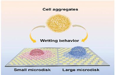
Cell aggregates as a 3D culture model can effectively mimic the physiological processes such as embryonic development, immune response, and tissue renewal in vivo. Researches show that the topography of biomaterials plays an important role in regulating cell proliferation, adhesion, and differentiation. It is of great significance to understand how cell aggregates respond to surface topography. Herein, microdisk array structures with the optimized size are used to investigate the wetting of cell aggregates. Cell aggregates exhibit complete wetting with distinct wetting velocities on the microdisk array structures of different diameters. The wetting velocity of cell aggregates reaches a maximum of 293 µm h−1 on microdisk structures with a diameter of 2 µm and is a minimum of 247 µm h−1 on microdisk structures of 20 µm diameter, which suggests that the cell-substrates adhesion energy on the latter is smaller. Actin stress fibers, focal adhesions (FAs), and cell morphology are analyzed to reveal the mechanisms of variation of wetting velocity. Furthermore, it is demonstrated that cell aggregates adopt climb and detour wetting modes on small and large-sized microdisk structures, respectively. This work reveals the response of cell aggregates to micro-scale topography, providing guidance for better understanding of tissue infiltration.
期刊介绍:
Small serves as an exceptional platform for both experimental and theoretical studies in fundamental and applied interdisciplinary research at the nano- and microscale. The journal offers a compelling mix of peer-reviewed Research Articles, Reviews, Perspectives, and Comments.
With a remarkable 2022 Journal Impact Factor of 13.3 (Journal Citation Reports from Clarivate Analytics, 2023), Small remains among the top multidisciplinary journals, covering a wide range of topics at the interface of materials science, chemistry, physics, engineering, medicine, and biology.
Small's readership includes biochemists, biologists, biomedical scientists, chemists, engineers, information technologists, materials scientists, physicists, and theoreticians alike.


 分享
分享
 求助内容:
求助内容: 应助结果提醒方式:
 扫码关注我们
