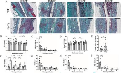
{"title":"在小鼠骨折修复过程中,成骨细胞 GαS 的增加通过抑制软骨和增强胼胝体矿化促进骨化","authors":"Kathy K Lee, Adele Changoor, Marc D Grynpas, Jane Mitchell","doi":"10.1002/jbm4.10841","DOIUrl":null,"url":null,"abstract":"<p>Gα<sub>S</sub>, the stimulatory G protein α-subunit that raises intracellular cAMP levels by activating adenylyl cyclase, plays a vital role in bone development, maintenance, and remodeling. Previously, using transgenic mice overexpressing Gα<sub>S</sub> in osteoblasts (G<sub>S</sub>-Tg), we demonstrated the influence of osteoblast Gα<sub>S</sub> level on osteogenesis, bone turnover, and skeletal responses to hyperparathyroidism. To further investigate whether alterations in Gα<sub>S</sub> levels affect endochondral bone repair, a postnatal bone regenerative process that recapitulates embryonic bone development, we performed stabilized tibial osteotomy in male G<sub>S</sub>-Tg mice at 8 weeks of age and examined the progression of fracture healing by micro-CT, histomorphometry, and gene expression analysis over a 4-week period. Bone fractures from G<sub>S</sub>-Tg mice exhibited diminished cartilage formation at the time of peak soft callus formation at 1 week post-fracture followed by significantly enhanced callus mineralization and new bone formation at 2 weeks post-fracture. The opposing effects on chondrogenesis and osteogenesis were validated by downregulation of chondrogenic markers and upregulation of osteogenic markers. Histomorphometric analysis at times of increased bone formation (2 and 3 weeks post-fracture) revealed excess fibroblast-like cells on newly formed woven bone surfaces and elevated osteocyte density in G<sub>S</sub>-Tg fractures. Coincident with enhanced callus mineralization and bone formation, G<sub>S</sub>-Tg mice showed elevated active β-catenin and Wntless proteins in osteoblasts at 2 weeks post-fracture, further substantiated by increased mRNA encoding various canonical Wnts and Wnt target genes, suggesting elevated osteoblastic Wnt secretion and Wnt/β-catenin signaling. The G<sub>S</sub>-Tg bony callus at 4 weeks post-fracture exhibited greater mineral density and decreased polar moment of inertia, resulting in improved material stiffness. These findings highlight that elevated Gα<sub>S</sub> levels increase Wnt signaling, conferring an increased osteogenic differentiation potential at the expense of chondrogenic differentiation, resulting in improved mechanical integrity. © 2023 The Authors. <i>JBMR Plus</i> published by Wiley Periodicals LLC. on behalf of American Society for Bone and Mineral Research.</p>","PeriodicalId":14611,"journal":{"name":"JBMR Plus","volume":"7 12","pages":""},"PeriodicalIF":3.4000,"publicationDate":"2023-11-15","publicationTypes":"Journal Article","fieldsOfStudy":null,"isOpenAccess":false,"openAccessPdf":"https://asbmr.onlinelibrary.wiley.com/doi/epdf/10.1002/jbm4.10841","citationCount":"0","resultStr":"{\"title\":\"Increased Osteoblast GαS Promotes Ossification by Suppressing Cartilage and Enhancing Callus Mineralization During Fracture Repair in Mice\",\"authors\":\"Kathy K Lee, Adele Changoor, Marc D Grynpas, Jane Mitchell\",\"doi\":\"10.1002/jbm4.10841\",\"DOIUrl\":null,\"url\":null,\"abstract\":\"<p>Gα<sub>S</sub>, the stimulatory G protein α-subunit that raises intracellular cAMP levels by activating adenylyl cyclase, plays a vital role in bone development, maintenance, and remodeling. Previously, using transgenic mice overexpressing Gα<sub>S</sub> in osteoblasts (G<sub>S</sub>-Tg), we demonstrated the influence of osteoblast Gα<sub>S</sub> level on osteogenesis, bone turnover, and skeletal responses to hyperparathyroidism. To further investigate whether alterations in Gα<sub>S</sub> levels affect endochondral bone repair, a postnatal bone regenerative process that recapitulates embryonic bone development, we performed stabilized tibial osteotomy in male G<sub>S</sub>-Tg mice at 8 weeks of age and examined the progression of fracture healing by micro-CT, histomorphometry, and gene expression analysis over a 4-week period. Bone fractures from G<sub>S</sub>-Tg mice exhibited diminished cartilage formation at the time of peak soft callus formation at 1 week post-fracture followed by significantly enhanced callus mineralization and new bone formation at 2 weeks post-fracture. The opposing effects on chondrogenesis and osteogenesis were validated by downregulation of chondrogenic markers and upregulation of osteogenic markers. Histomorphometric analysis at times of increased bone formation (2 and 3 weeks post-fracture) revealed excess fibroblast-like cells on newly formed woven bone surfaces and elevated osteocyte density in G<sub>S</sub>-Tg fractures. Coincident with enhanced callus mineralization and bone formation, G<sub>S</sub>-Tg mice showed elevated active β-catenin and Wntless proteins in osteoblasts at 2 weeks post-fracture, further substantiated by increased mRNA encoding various canonical Wnts and Wnt target genes, suggesting elevated osteoblastic Wnt secretion and Wnt/β-catenin signaling. The G<sub>S</sub>-Tg bony callus at 4 weeks post-fracture exhibited greater mineral density and decreased polar moment of inertia, resulting in improved material stiffness. These findings highlight that elevated Gα<sub>S</sub> levels increase Wnt signaling, conferring an increased osteogenic differentiation potential at the expense of chondrogenic differentiation, resulting in improved mechanical integrity. © 2023 The Authors. <i>JBMR Plus</i> published by Wiley Periodicals LLC. on behalf of American Society for Bone and Mineral Research.</p>\",\"PeriodicalId\":14611,\"journal\":{\"name\":\"JBMR Plus\",\"volume\":\"7 12\",\"pages\":\"\"},\"PeriodicalIF\":3.4000,\"publicationDate\":\"2023-11-15\",\"publicationTypes\":\"Journal Article\",\"fieldsOfStudy\":null,\"isOpenAccess\":false,\"openAccessPdf\":\"https://asbmr.onlinelibrary.wiley.com/doi/epdf/10.1002/jbm4.10841\",\"citationCount\":\"0\",\"resultStr\":null,\"platform\":\"Semanticscholar\",\"paperid\":null,\"PeriodicalName\":\"JBMR Plus\",\"FirstCategoryId\":\"1085\",\"ListUrlMain\":\"https://onlinelibrary.wiley.com/doi/10.1002/jbm4.10841\",\"RegionNum\":0,\"RegionCategory\":null,\"ArticlePicture\":[],\"TitleCN\":null,\"AbstractTextCN\":null,\"PMCID\":null,\"EPubDate\":\"\",\"PubModel\":\"\",\"JCR\":\"Q2\",\"JCRName\":\"ENDOCRINOLOGY & METABOLISM\",\"Score\":null,\"Total\":0}","platform":"Semanticscholar","paperid":null,"PeriodicalName":"JBMR Plus","FirstCategoryId":"1085","ListUrlMain":"https://onlinelibrary.wiley.com/doi/10.1002/jbm4.10841","RegionNum":0,"RegionCategory":null,"ArticlePicture":[],"TitleCN":null,"AbstractTextCN":null,"PMCID":null,"EPubDate":"","PubModel":"","JCR":"Q2","JCRName":"ENDOCRINOLOGY & METABOLISM","Score":null,"Total":0}
引用次数: 0


