Apurva Singh, Leonid Roshkovan, Hannah Horng, Andrew Chen, Sharyn I Katz, Jeffrey C Thompson, Despina Kontos
{"title":"从纵向手术前 CT 扫描中识别侵袭性肺实性下结节的放射组学分析","authors":"Apurva Singh, Leonid Roshkovan, Hannah Horng, Andrew Chen, Sharyn I Katz, Jeffrey C Thompson, Despina Kontos","doi":"10.1097/RTI.0000000000000800","DOIUrl":null,"url":null,"abstract":"<p><strong>Purpose: </strong>Effective identification of malignant part-solid lung nodules is crucial to eliminate risks due to therapeutic intervention or lack thereof. We aimed to develop delta radiomics and volumetric signatures, characterize changes in nodule properties over three presurgical time points, and assess the accuracy of nodule invasiveness identification when combined with immediate presurgical time point radiomics signature and clinical biomarkers.</p><p><strong>Materials and methods: </strong>Cohort included 156 part-solid lung nodules with immediate presurgical CT scans and a subset of 122 nodules with scans at 3 presurgical time points. Region of interest segmentation was performed using ITK-SNAP, and feature extraction using CaPTk. Image parameter heterogeneity was mitigated at each time point using nested ComBat harmonization. For 122 nodules, delta radiomics features (ΔR AB = (R B -R A )/R A ) and delta volumes (ΔV AB = (V B -V A )/V A ) were computed between the time points. Principal Component Analysis was performed to construct immediate presurgical radiomics (Rs 1 ) and delta radiomics signatures (ΔRs 31 + ΔRs 21 + ΔRs 32 ). Identification of nodule pathology was performed using logistic regression on delta radiomics and immediate presurgical time point signatures, delta volumes (ΔV 31 + ΔV 21 + ΔV 32 ), and clinical variable (smoking status, BMI) models (train test split (2:1)).</p><p><strong>Results: </strong>In delta radiomics analysis (n= 122 nodules), the best-performing model combined immediate pre-surgical time point and delta radiomics signatures, delta volumes, and clinical factors (classification accuracy [AUC]): (77.5% [0.73]) (train); (71.6% [0.69]) (test).</p><p><strong>Conclusions: </strong>Delta radiomics and volumes can detect changes in nodule properties over time, which are predictive of nodule invasiveness. These tools could improve conventional radiologic assessment, allow for earlier intervention for aggressive nodules, and decrease unnecessary intervention-related morbidity.</p>","PeriodicalId":49974,"journal":{"name":"Journal of Thoracic Imaging","volume":" ","pages":""},"PeriodicalIF":1.9000,"publicationDate":"2025-01-01","publicationTypes":"Journal Article","fieldsOfStudy":null,"isOpenAccess":false,"openAccessPdf":"https://www.ncbi.nlm.nih.gov/pmc/articles/PMC11654445/pdf/","citationCount":"0","resultStr":"{\"title\":\"Radiomics Analysis for the Identification of Invasive Pulmonary Subsolid Nodules From Longitudinal Presurgical CT Scans.\",\"authors\":\"Apurva Singh, Leonid Roshkovan, Hannah Horng, Andrew Chen, Sharyn I Katz, Jeffrey C Thompson, Despina Kontos\",\"doi\":\"10.1097/RTI.0000000000000800\",\"DOIUrl\":null,\"url\":null,\"abstract\":\"<p><strong>Purpose: </strong>Effective identification of malignant part-solid lung nodules is crucial to eliminate risks due to therapeutic intervention or lack thereof. We aimed to develop delta radiomics and volumetric signatures, characterize changes in nodule properties over three presurgical time points, and assess the accuracy of nodule invasiveness identification when combined with immediate presurgical time point radiomics signature and clinical biomarkers.</p><p><strong>Materials and methods: </strong>Cohort included 156 part-solid lung nodules with immediate presurgical CT scans and a subset of 122 nodules with scans at 3 presurgical time points. Region of interest segmentation was performed using ITK-SNAP, and feature extraction using CaPTk. Image parameter heterogeneity was mitigated at each time point using nested ComBat harmonization. For 122 nodules, delta radiomics features (ΔR AB = (R B -R A )/R A ) and delta volumes (ΔV AB = (V B -V A )/V A ) were computed between the time points. Principal Component Analysis was performed to construct immediate presurgical radiomics (Rs 1 ) and delta radiomics signatures (ΔRs 31 + ΔRs 21 + ΔRs 32 ). Identification of nodule pathology was performed using logistic regression on delta radiomics and immediate presurgical time point signatures, delta volumes (ΔV 31 + ΔV 21 + ΔV 32 ), and clinical variable (smoking status, BMI) models (train test split (2:1)).</p><p><strong>Results: </strong>In delta radiomics analysis (n= 122 nodules), the best-performing model combined immediate pre-surgical time point and delta radiomics signatures, delta volumes, and clinical factors (classification accuracy [AUC]): (77.5% [0.73]) (train); (71.6% [0.69]) (test).</p><p><strong>Conclusions: </strong>Delta radiomics and volumes can detect changes in nodule properties over time, which are predictive of nodule invasiveness. These tools could improve conventional radiologic assessment, allow for earlier intervention for aggressive nodules, and decrease unnecessary intervention-related morbidity.</p>\",\"PeriodicalId\":49974,\"journal\":{\"name\":\"Journal of Thoracic Imaging\",\"volume\":\" \",\"pages\":\"\"},\"PeriodicalIF\":1.9000,\"publicationDate\":\"2025-01-01\",\"publicationTypes\":\"Journal Article\",\"fieldsOfStudy\":null,\"isOpenAccess\":false,\"openAccessPdf\":\"https://www.ncbi.nlm.nih.gov/pmc/articles/PMC11654445/pdf/\",\"citationCount\":\"0\",\"resultStr\":null,\"platform\":\"Semanticscholar\",\"paperid\":null,\"PeriodicalName\":\"Journal of Thoracic Imaging\",\"FirstCategoryId\":\"3\",\"ListUrlMain\":\"https://doi.org/10.1097/RTI.0000000000000800\",\"RegionNum\":4,\"RegionCategory\":\"医学\",\"ArticlePicture\":[],\"TitleCN\":null,\"AbstractTextCN\":null,\"PMCID\":null,\"EPubDate\":\"\",\"PubModel\":\"\",\"JCR\":\"Q3\",\"JCRName\":\"RADIOLOGY, NUCLEAR MEDICINE & MEDICAL IMAGING\",\"Score\":null,\"Total\":0}","platform":"Semanticscholar","paperid":null,"PeriodicalName":"Journal of Thoracic Imaging","FirstCategoryId":"3","ListUrlMain":"https://doi.org/10.1097/RTI.0000000000000800","RegionNum":4,"RegionCategory":"医学","ArticlePicture":[],"TitleCN":null,"AbstractTextCN":null,"PMCID":null,"EPubDate":"","PubModel":"","JCR":"Q3","JCRName":"RADIOLOGY, NUCLEAR MEDICINE & MEDICAL IMAGING","Score":null,"Total":0}
Radiomics Analysis for the Identification of Invasive Pulmonary Subsolid Nodules From Longitudinal Presurgical CT Scans.
Purpose: Effective identification of malignant part-solid lung nodules is crucial to eliminate risks due to therapeutic intervention or lack thereof. We aimed to develop delta radiomics and volumetric signatures, characterize changes in nodule properties over three presurgical time points, and assess the accuracy of nodule invasiveness identification when combined with immediate presurgical time point radiomics signature and clinical biomarkers.
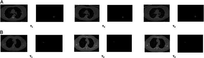
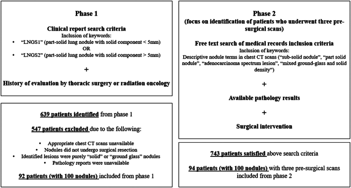
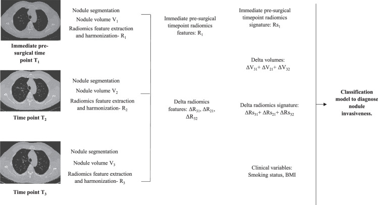
Materials and methods: Cohort included 156 part-solid lung nodules with immediate presurgical CT scans and a subset of 122 nodules with scans at 3 presurgical time points. Region of interest segmentation was performed using ITK-SNAP, and feature extraction using CaPTk. Image parameter heterogeneity was mitigated at each time point using nested ComBat harmonization. For 122 nodules, delta radiomics features (ΔR AB = (R B -R A )/R A ) and delta volumes (ΔV AB = (V B -V A )/V A ) were computed between the time points. Principal Component Analysis was performed to construct immediate presurgical radiomics (Rs 1 ) and delta radiomics signatures (ΔRs 31 + ΔRs 21 + ΔRs 32 ). Identification of nodule pathology was performed using logistic regression on delta radiomics and immediate presurgical time point signatures, delta volumes (ΔV 31 + ΔV 21 + ΔV 32 ), and clinical variable (smoking status, BMI) models (train test split (2:1)).
Results: In delta radiomics analysis (n= 122 nodules), the best-performing model combined immediate pre-surgical time point and delta radiomics signatures, delta volumes, and clinical factors (classification accuracy [AUC]): (77.5% [0.73]) (train); (71.6% [0.69]) (test).
Conclusions: Delta radiomics and volumes can detect changes in nodule properties over time, which are predictive of nodule invasiveness. These tools could improve conventional radiologic assessment, allow for earlier intervention for aggressive nodules, and decrease unnecessary intervention-related morbidity.
期刊介绍:
Journal of Thoracic Imaging (JTI) provides authoritative information on all aspects of the use of imaging techniques in the diagnosis of cardiac and pulmonary diseases. Original articles and analytical reviews published in this timely journal provide the very latest thinking of leading experts concerning the use of chest radiography, computed tomography, magnetic resonance imaging, positron emission tomography, ultrasound, and all other promising imaging techniques in cardiopulmonary radiology.
Official Journal of the Society of Thoracic Radiology:
Japanese Society of Thoracic Radiology
Korean Society of Thoracic Radiology
European Society of Thoracic Imaging.


 分享
分享
 求助内容:
求助内容: 应助结果提醒方式:
 扫码关注我们
