Reina Fujiwara, Kie Yamamoto, Masahiro Yamasaki, Koichi Ohno
{"title":"Magnetic Resonance Cholangiopancreatography for Cholangiopancreatic Duct Imaging in Dogs.","authors":"Reina Fujiwara, Kie Yamamoto, Masahiro Yamasaki, Koichi Ohno","doi":"10.1111/vru.70008","DOIUrl":null,"url":null,"abstract":"<p><p>Ultrasonography is often used to diagnose biliary diseases in dogs; however, it is difficult to delineate the entire bile and pancreatic ducts. Various imaging techniques for bile and pancreatic ducts have been attempted to overcome this problem. Magnetic resonance cholangiopancreatography (MRCP) is often used to evaluate the bile and pancreatic ducts in humans with obstructive jaundice, but very few reports exist on its usage in dogs. This study was designed as a prospective observational study to assess the feasibility and effectiveness of MRCP for visualizing the bile and pancreatic ducts in nondiseased dogs. Therefore, this study aimed to evaluate the visibility of the bile and pancreatic ducts through MRCP imaging using a 3.0 T-MRI system in dogs with no signs of hepatic, biliary, and pancreatic diseases. The detection rate for each anatomical structure was evaluated, with the highest observed in the gallbladder (100%), followed by the common bile duct (80%), cystic duct (70%), pancreatic ducts in the left and right lobe of the pancreas (70%), left and right hepatic ducts (60%), accessory pancreatic ducts (60%), and major pancreatic duct (40%). MRCP is a promising noninvasive imaging technique that can promptly and accurately visualize bile and pancreatic ducts in dogs without being influenced by the skill of the operator.</p>","PeriodicalId":23581,"journal":{"name":"Veterinary Radiology & Ultrasound","volume":"66 1","pages":"e70008"},"PeriodicalIF":1.5000,"publicationDate":"2025-01-01","publicationTypes":"Journal Article","fieldsOfStudy":null,"isOpenAccess":false,"openAccessPdf":"https://www.ncbi.nlm.nih.gov/pmc/articles/PMC11742707/pdf/","citationCount":"0","resultStr":null,"platform":"Semanticscholar","paperid":null,"PeriodicalName":"Veterinary Radiology & Ultrasound","FirstCategoryId":"97","ListUrlMain":"https://doi.org/10.1111/vru.70008","RegionNum":2,"RegionCategory":"农林科学","ArticlePicture":[],"TitleCN":null,"AbstractTextCN":null,"PMCID":null,"EPubDate":"","PubModel":"","JCR":"Q2","JCRName":"VETERINARY SCIENCES","Score":null,"Total":0}
引用次数: 0
Abstract
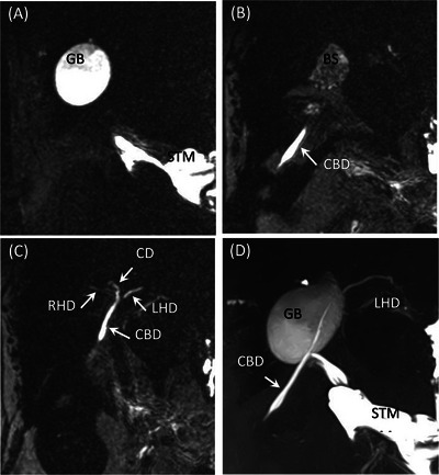
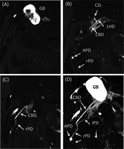
Ultrasonography is often used to diagnose biliary diseases in dogs; however, it is difficult to delineate the entire bile and pancreatic ducts. Various imaging techniques for bile and pancreatic ducts have been attempted to overcome this problem. Magnetic resonance cholangiopancreatography (MRCP) is often used to evaluate the bile and pancreatic ducts in humans with obstructive jaundice, but very few reports exist on its usage in dogs. This study was designed as a prospective observational study to assess the feasibility and effectiveness of MRCP for visualizing the bile and pancreatic ducts in nondiseased dogs. Therefore, this study aimed to evaluate the visibility of the bile and pancreatic ducts through MRCP imaging using a 3.0 T-MRI system in dogs with no signs of hepatic, biliary, and pancreatic diseases. The detection rate for each anatomical structure was evaluated, with the highest observed in the gallbladder (100%), followed by the common bile duct (80%), cystic duct (70%), pancreatic ducts in the left and right lobe of the pancreas (70%), left and right hepatic ducts (60%), accessory pancreatic ducts (60%), and major pancreatic duct (40%). MRCP is a promising noninvasive imaging technique that can promptly and accurately visualize bile and pancreatic ducts in dogs without being influenced by the skill of the operator.
期刊介绍:
Veterinary Radiology & Ultrasound is a bimonthly, international, peer-reviewed, research journal devoted to the fields of veterinary diagnostic imaging and radiation oncology. Established in 1958, it is owned by the American College of Veterinary Radiology and is also the official journal for six affiliate veterinary organizations. Veterinary Radiology & Ultrasound is represented on the International Committee of Medical Journal Editors, World Association of Medical Editors, and Committee on Publication Ethics.
The mission of Veterinary Radiology & Ultrasound is to serve as a leading resource for high quality articles that advance scientific knowledge and standards of clinical practice in the areas of veterinary diagnostic radiology, computed tomography, magnetic resonance imaging, ultrasonography, nuclear imaging, radiation oncology, and interventional radiology. Manuscript types include original investigations, imaging diagnosis reports, review articles, editorials and letters to the Editor. Acceptance criteria include originality, significance, quality, reader interest, composition and adherence to author guidelines.


 分享
分享
 求助内容:
求助内容: 应助结果提醒方式:
 扫码关注我们
