Pub Date : 2024-06-01Epub Date: 2024-03-21DOI: 10.1262/jrd.2024-005
Hiromi Kusaka, Minoru Sakaguchi
The number of cows in estrus often influences estrus behavior; however, the effects of social order are not well documented. This study examined the effects of social order on the expression of behaviorally-scored and pedometer-detected estrus, combined with the effects of the number of cows in estrus. In a herd comprising 13 or 15 beef cattle, cows with orders 1st-7th were defined as dominant and the remaining cows as subordinate. Sole or simultaneous estrus was induced by prostaglandin F2α analog injection and/or intravaginal progesterone treatment. Ovulation timing was determined using ultrasonography at 6-hour intervals. Estrous signs and steps of the cows were recorded 49 h before ovulation using video monitoring and a pedometer, respectively. Among the 59 treated cows, 56 behaviorally-scored estruses (27 sole and 29 simultaneous) were detected. In the sole estrus, 61.5% of the dominant-rank cows had no zero-point period; however, 35.7% of the subordinate-rank cows had that period. The dominant-rank cows in estrus alone had a significantly shorter duration of scored estrus than those in simultaneous estrus (P < 0.05). Among the 50 pedometer-detected estruses (24 sole and 26 simultaneous), the subordinate-rank cows in sole estrus had a shorter interval from estrus onset to ovulation than the dominant-rank cows in simultaneous estrus (P < 0.05). The effects of social order varied in response to the number of cows in estrus, which might have influenced determining the optimal time for artificial insemination.
{"title":"Social effects on behaviorally-scored and pedometer-detected estrus in beef cattle.","authors":"Hiromi Kusaka, Minoru Sakaguchi","doi":"10.1262/jrd.2024-005","DOIUrl":"10.1262/jrd.2024-005","url":null,"abstract":"<p><p>The number of cows in estrus often influences estrus behavior; however, the effects of social order are not well documented. This study examined the effects of social order on the expression of behaviorally-scored and pedometer-detected estrus, combined with the effects of the number of cows in estrus. In a herd comprising 13 or 15 beef cattle, cows with orders 1st-7th were defined as dominant and the remaining cows as subordinate. Sole or simultaneous estrus was induced by prostaglandin F<sub>2α</sub> analog injection and/or intravaginal progesterone treatment. Ovulation timing was determined using ultrasonography at 6-hour intervals. Estrous signs and steps of the cows were recorded 49 h before ovulation using video monitoring and a pedometer, respectively. Among the 59 treated cows, 56 behaviorally-scored estruses (27 sole and 29 simultaneous) were detected. In the sole estrus, 61.5% of the dominant-rank cows had no zero-point period; however, 35.7% of the subordinate-rank cows had that period. The dominant-rank cows in estrus alone had a significantly shorter duration of scored estrus than those in simultaneous estrus (P < 0.05). Among the 50 pedometer-detected estruses (24 sole and 26 simultaneous), the subordinate-rank cows in sole estrus had a shorter interval from estrus onset to ovulation than the dominant-rank cows in simultaneous estrus (P < 0.05). The effects of social order varied in response to the number of cows in estrus, which might have influenced determining the optimal time for artificial insemination.</p>","PeriodicalId":16942,"journal":{"name":"Journal of Reproduction and Development","volume":" ","pages":"177-183"},"PeriodicalIF":1.8,"publicationDate":"2024-06-01","publicationTypes":"Journal Article","fieldsOfStudy":null,"isOpenAccess":false,"openAccessPdf":"https://www.ncbi.nlm.nih.gov/pmc/articles/PMC11153121/pdf/","citationCount":null,"resultStr":null,"platform":"Semanticscholar","paperid":"140175075","PeriodicalName":null,"FirstCategoryId":null,"ListUrlMain":null,"RegionNum":4,"RegionCategory":"生物学","ArticlePicture":[],"TitleCN":null,"AbstractTextCN":null,"PMCID":"OA","EPubDate":null,"PubModel":null,"JCR":null,"JCRName":null,"Score":null,"Total":0}
Ovarian stimulation protocols are widely used to collect oocytes in assisted reproductive technologies (ARTs). Although the influence of ovarian stimulation on embryo quality has been described, this issue remains controversial. Here, we analyzed the influence of ovarian stimulation on developmental speed and chromosome segregation using live cell imaging. Female mice at the proestrus stage were separated by the appearance of the vagina as the non-stimulation (-) group, and other mice were administered pregnant mare serum gonadotropin (PMSG) and human chorionic gonadotropin (hCG) as the stimulation (+) groups. The cumulus-oocyte complexes from both groups were inseminated with sperm suspensions from the same male mice. Fertilization rates and developmental capacities were examined, and the developmental speed and frequency of chromosome segregation errors were measured by live-cell imaging using a Histone H2B-mCherry probe. The number of fertilized oocytes obtained was 1.4-fold more frequent in the stimulation (+) group. The developmental rate and chromosome stability did not differ between the groups. Image analysis showed that the mean speed of development in the stimulation (+) group was slightly higher than that in the non-stimulation (-) group. This increase in speed seemed to arise from the slight shortening of the 2- and 4-cell stages and third division lengths and consequent synchronization of cleavage timing in each embryo, not from the emergence of an extremely rapidly developing subpopulation of embryos. In conclusion, ovarian stimulation does not necessarily affect embryo quality but rather increases the chances of obtaining high-quality oocytes in mice.
{"title":"Effect of ovarian stimulation on developmental speed of preimplantation embryo in a mouse model.","authors":"Mayuko Kurumizaka, Tatsuma Yao, Mikiko Tokoro, Noritaka Fukunaga, Yoshimasa Asada, Kazuo Yamagata","doi":"10.1262/jrd.2023-089","DOIUrl":"10.1262/jrd.2023-089","url":null,"abstract":"<p><p>Ovarian stimulation protocols are widely used to collect oocytes in assisted reproductive technologies (ARTs). Although the influence of ovarian stimulation on embryo quality has been described, this issue remains controversial. Here, we analyzed the influence of ovarian stimulation on developmental speed and chromosome segregation using live cell imaging. Female mice at the proestrus stage were separated by the appearance of the vagina as the non-stimulation (-) group, and other mice were administered pregnant mare serum gonadotropin (PMSG) and human chorionic gonadotropin (hCG) as the stimulation (+) groups. The cumulus-oocyte complexes from both groups were inseminated with sperm suspensions from the same male mice. Fertilization rates and developmental capacities were examined, and the developmental speed and frequency of chromosome segregation errors were measured by live-cell imaging using a Histone H2B-mCherry probe. The number of fertilized oocytes obtained was 1.4-fold more frequent in the stimulation (+) group. The developmental rate and chromosome stability did not differ between the groups. Image analysis showed that the mean speed of development in the stimulation (+) group was slightly higher than that in the non-stimulation (-) group. This increase in speed seemed to arise from the slight shortening of the 2- and 4-cell stages and third division lengths and consequent synchronization of cleavage timing in each embryo, not from the emergence of an extremely rapidly developing subpopulation of embryos. In conclusion, ovarian stimulation does not necessarily affect embryo quality but rather increases the chances of obtaining high-quality oocytes in mice.</p>","PeriodicalId":16942,"journal":{"name":"Journal of Reproduction and Development","volume":" ","pages":"160-168"},"PeriodicalIF":1.8,"publicationDate":"2024-06-01","publicationTypes":"Journal Article","fieldsOfStudy":null,"isOpenAccess":false,"openAccessPdf":"https://www.ncbi.nlm.nih.gov/pmc/articles/PMC11153123/pdf/","citationCount":null,"resultStr":null,"platform":"Semanticscholar","paperid":"140143663","PeriodicalName":null,"FirstCategoryId":null,"ListUrlMain":null,"RegionNum":4,"RegionCategory":"生物学","ArticlePicture":[],"TitleCN":null,"AbstractTextCN":null,"PMCID":"OA","EPubDate":null,"PubModel":null,"JCR":null,"JCRName":null,"Score":null,"Total":0}
Mammalian reproduction is more inefficient than expected and embryo/conceptus implantation into the maternal endometrium is considered to be a rate-limiting process. Although extensive physiological and structural diversity exists among mammalian species, the basic molecular mechanisms underlying successful implantation are conserved. The extensive use of genetically engineered mouse models has provided considerable information on uterine receptivity for embryo implantation. The molecular mechanisms and cellular processes identified thus far require further validation in other mammalian species. In this review, representative ovarian steroid hormone-induced signaling pathways controlling uterine adaptation are presented based on the results of rodent studies. Selected examples of functional conservation in mammals, such as humans and cattle, are briefly described. To date, molecular therapeutic trials for fertility improvement have not been conducted. Considerable efforts are required to provide further understanding of these molecular mechanisms. Such understanding will contribute to the development of reliable clinical diagnostics and therapeutics for implantation failure, leading to reproductive success in a wide variety of mammals in the future.
{"title":"Molecular approaches to mammalian uterine receptivity for conceptus implantation","authors":"Kaiyu KUBOTA","doi":"10.1262/jrd.2024-022","DOIUrl":"https://doi.org/10.1262/jrd.2024-022","url":null,"abstract":"</p><p>Mammalian reproduction is more inefficient than expected and embryo/conceptus implantation into the maternal endometrium is considered to be a rate-limiting process. Although extensive physiological and structural diversity exists among mammalian species, the basic molecular mechanisms underlying successful implantation are conserved. The extensive use of genetically engineered mouse models has provided considerable information on uterine receptivity for embryo implantation. The molecular mechanisms and cellular processes identified thus far require further validation in other mammalian species. In this review, representative ovarian steroid hormone-induced signaling pathways controlling uterine adaptation are presented based on the results of rodent studies. Selected examples of functional conservation in mammals, such as humans and cattle, are briefly described. To date, molecular therapeutic trials for fertility improvement have not been conducted. Considerable efforts are required to provide further understanding of these molecular mechanisms. Such understanding will contribute to the development of reliable clinical diagnostics and therapeutics for implantation failure, leading to reproductive success in a wide variety of mammals in the future.</p>\u0000<p></p>\u0000<img alt=\"\" src=\"https://www.jstage.jst.go.jp/pub/jrd/advpub/0/advpub_2024-022/figure/advpub_2024-022.png\"/>\u0000Graphical Abstract <span style=\"padding-left:5px;\">Fullsize Image</span>","PeriodicalId":16942,"journal":{"name":"Journal of Reproduction and Development","volume":"50 1","pages":""},"PeriodicalIF":1.8,"publicationDate":"2024-05-18","publicationTypes":"Journal Article","fieldsOfStudy":null,"isOpenAccess":false,"openAccessPdf":"","citationCount":null,"resultStr":null,"platform":"Semanticscholar","paperid":"141064218","PeriodicalName":null,"FirstCategoryId":null,"ListUrlMain":null,"RegionNum":4,"RegionCategory":"生物学","ArticlePicture":[],"TitleCN":null,"AbstractTextCN":null,"PMCID":"","EPubDate":null,"PubModel":null,"JCR":null,"JCRName":null,"Score":null,"Total":0}
Embryonic transfer of bovine blastocysts produced by in vitro fertilization is widely utilized despite a compromised conception rate. It has been suggested that a set of four evaluation criteria for judging the quality of embryos, based on the timing of early cleavages and proper morphologies of embryos, can effectively predict pregnancy success. These blastocysts are hereafter referred to as four-criteria-compliant blastocysts. The same criteria should be used to modify the culture media to improve embryo quality. For example, culture media is often supplemented with nonessential amino acids (NEAA) at a uniform concentration despite the major variation in their concentration in the oviductal fluid. In the present study, the effects of the embryo culture medium, namely CR1, supplemented with all seven MEM NEAA or six of them, excluding one at a time, were examined. All media, except for the medium that did not contain proline and serine, tended to improve the efficiency of producing four-criteria-compliant blastocysts, and excluding alanine was particularly effective. The absence of alanine resulted in the rapid occurrence of the first cleavage and pronuclear formation of fertilized oocytes in the alanine-free medium compared to that in the medium containing alanine. These results suggested that alanine hinders certain events involved in the progression of early embryogenesis, which is necessary to achieve the four criteria that provide a benchmark for pregnancy. Therefore, a significantly higher percentage of embryos satisfied the recommended criteria and developed into four-criteria-compliant blastocysts when developed in alanine-free medium than in alanine-containing medium.
���Graphical Abstract Fullsize Image
尽管受孕率不高,但体外受精产生的牛囊胚的胚胎移植仍被广泛使用。有研究表明,根据胚胎早期分裂的时间和胚胎的适当形态来判断胚胎质量的一套四项评价标准可有效预测妊娠成功率。这些囊胚在下文中称为符合四项标准的囊胚。应使用相同的标准来修改培养基,以提高胚胎质量。例如,尽管非必需氨基酸(NEAA)在输卵管液中的浓度存在很大差异,但培养基中通常会添加统一浓度的非必需氨基酸。本研究考察了胚胎培养基(即 CR1)补充全部七种 MEM 非必需氨基酸或六种非必需氨基酸(每次排除一种)的效果。除了不含脯氨酸和丝氨酸的培养基外,所有培养基都倾向于提高生产符合四项标准的囊胚的效率,不含丙氨酸的培养基尤其有效。与含丙氨酸的培养基相比,不含丙氨酸的培养基中受精卵细胞的第一次分裂和原核形成更快。这些结果表明,丙氨酸阻碍了早期胚胎发生过程中的某些事件,而早期胚胎发生是达到怀孕基准的四项标准所必需的。因此,与含丙氨酸的培养基相比,在无丙氨酸培养基中发育的胚胎符合推荐标准并发育成符合四项标准的囊胚的比例要高得多。 图文摘要 全尺寸图片
{"title":"Excluding alanine from minimum essential medium (MEM) nonessential amino acid supplementation of the culture medium facilitates post-fertilization events and early cleavages of bovine oocytes fertilized in vitro","authors":"Nobuhiko ITAMI, Satoshi AKAGI, Yuji HIRAO","doi":"10.1262/jrd.2023-098","DOIUrl":"https://doi.org/10.1262/jrd.2023-098","url":null,"abstract":"</p><p>Embryonic transfer of bovine blastocysts produced by <i>in vitro</i> fertilization is widely utilized despite a compromised conception rate. It has been suggested that a set of four evaluation criteria for judging the quality of embryos, based on the timing of early cleavages and proper morphologies of embryos, can effectively predict pregnancy success. These blastocysts are hereafter referred to as four-criteria-compliant blastocysts. The same criteria should be used to modify the culture media to improve embryo quality. For example, culture media is often supplemented with nonessential amino acids (NEAA) at a uniform concentration despite the major variation in their concentration in the oviductal fluid. In the present study, the effects of the embryo culture medium, namely CR1, supplemented with all seven MEM NEAA or six of them, excluding one at a time, were examined. All media, except for the medium that did not contain proline and serine, tended to improve the efficiency of producing four-criteria-compliant blastocysts, and excluding alanine was particularly effective. The absence of alanine resulted in the rapid occurrence of the first cleavage and pronuclear formation of fertilized oocytes in the alanine-free medium compared to that in the medium containing alanine. These results suggested that alanine hinders certain events involved in the progression of early embryogenesis, which is necessary to achieve the four criteria that provide a benchmark for pregnancy. Therefore, a significantly higher percentage of embryos satisfied the recommended criteria and developed into four-criteria-compliant blastocysts when developed in alanine-free medium than in alanine-containing medium.</p>\u0000<p></p>\u0000<img alt=\"\" src=\"https://www.jstage.jst.go.jp/pub/jrd/advpub/0/advpub_2023-098/figure/advpub_2023-098.png\"/>\u0000Graphical Abstract <span style=\"padding-left:5px;\">Fullsize Image</span>","PeriodicalId":16942,"journal":{"name":"Journal of Reproduction and Development","volume":"48 1","pages":""},"PeriodicalIF":1.8,"publicationDate":"2024-05-17","publicationTypes":"Journal Article","fieldsOfStudy":null,"isOpenAccess":false,"openAccessPdf":"","citationCount":null,"resultStr":null,"platform":"Semanticscholar","paperid":"141058901","PeriodicalName":null,"FirstCategoryId":null,"ListUrlMain":null,"RegionNum":4,"RegionCategory":"生物学","ArticlePicture":[],"TitleCN":null,"AbstractTextCN":null,"PMCID":"","EPubDate":null,"PubModel":null,"JCR":null,"JCRName":null,"Score":null,"Total":0}
Understanding of central nervous system mechanisms underlying age-related infertility remains limited. Fibril α-synuclein, distinct from its monomeric form, is implicated in age-related diseases. Notably, fibril α-synuclein spreads among neurons, similar to prions, from damaged old neurons in cortex and hippocampus to healthy neurons. However, less is known whether α-synuclein propagates into oxytocin neurons, which play crucial roles in reproduction. We compared α-synuclein expression in the oxytocin neurons in suprachiasmatic nucleus (SCN), supraoptic nucleus (SON), paraventricular hypothalamic nucleus (PVN), and posterior pituitary (PP) gland of healthy heifers and aged cows to determine its role in age-related infertility. We analyzed mRNA and protein expression, along with Congo red histochemistry and fluorescent immunohistochemistry for oxytocin and α-synuclein, followed by confocal microscopy with Congo red staining. Both mRNA and protein expressions of α-synuclein were confirmed in the bovine cortex, hippocampus, SCN, SON, PVN, and PP tissues. Significant differences in α-synuclein mRNA expressions were observed in the cortex and hippocampus between young heifers and old cows. Western blots showed five bands of α-synuclein, probably reflecting monomers, dimers, and oligomers, in the cortex, hippocampus, SCN, SON, PVN, and PP tissues, and there were significant differences in some bands between the young heifers and old cows. Bright-field and polarized light microscopy did not detect obvious amyloid deposition in the aged hypothalami; however, higher-sensitive confocal microscopy unveiled strong positive signals for Congo red and α-synuclein in oxytocin neurons in the aged hypothalami. α-synuclein was expressed in oxytocin neurons, and some differences were observed between young and old hypothalami.
{"title":"Alpha-synuclein expression in oxytocin neurons of young and old bovine brains","authors":"Yvan Bienvenu NIYONZIMA, Yuuki ASATO, Hiroya KADOKAWA","doi":"10.1262/jrd.2024-020","DOIUrl":"https://doi.org/10.1262/jrd.2024-020","url":null,"abstract":"</p><p>Understanding of central nervous system mechanisms underlying age-related infertility remains limited. Fibril α-synuclein, distinct from its monomeric form, is implicated in age-related diseases. Notably, fibril α-synuclein spreads among neurons, similar to prions, from damaged old neurons in cortex and hippocampus to healthy neurons. However, less is known whether α-synuclein propagates into oxytocin neurons, which play crucial roles in reproduction. We compared α-synuclein expression in the oxytocin neurons in suprachiasmatic nucleus (SCN), supraoptic nucleus (SON), paraventricular hypothalamic nucleus (PVN), and posterior pituitary (PP) gland of healthy heifers and aged cows to determine its role in age-related infertility. We analyzed mRNA and protein expression, along with Congo red histochemistry and fluorescent immunohistochemistry for oxytocin and α-synuclein, followed by confocal microscopy with Congo red staining. Both mRNA and protein expressions of α-synuclein were confirmed in the bovine cortex, hippocampus, SCN, SON, PVN, and PP tissues. Significant differences in α-synuclein mRNA expressions were observed in the cortex and hippocampus between young heifers and old cows. Western blots showed five bands of α-synuclein, probably reflecting monomers, dimers, and oligomers, in the cortex, hippocampus, SCN, SON, PVN, and PP tissues, and there were significant differences in some bands between the young heifers and old cows. Bright-field and polarized light microscopy did not detect obvious amyloid deposition in the aged hypothalami; however, higher-sensitive confocal microscopy unveiled strong positive signals for Congo red and α-synuclein in oxytocin neurons in the aged hypothalami. α-synuclein was expressed in oxytocin neurons, and some differences were observed between young and old hypothalami.</p>\u0000<p></p>\u0000<img alt=\"\" src=\"https://www.jstage.jst.go.jp/pub/jrd/advpub/0/advpub_2024-020/figure/advpub_2024-020.jpg\"/>\u0000Graphical Abstract <span style=\"padding-left:5px;\">Fullsize Image</span>","PeriodicalId":16942,"journal":{"name":"Journal of Reproduction and Development","volume":"129 1","pages":""},"PeriodicalIF":1.8,"publicationDate":"2024-04-29","publicationTypes":"Journal Article","fieldsOfStudy":null,"isOpenAccess":false,"openAccessPdf":"","citationCount":null,"resultStr":null,"platform":"Semanticscholar","paperid":"140809370","PeriodicalName":null,"FirstCategoryId":null,"ListUrlMain":null,"RegionNum":4,"RegionCategory":"生物学","ArticlePicture":[],"TitleCN":null,"AbstractTextCN":null,"PMCID":"","EPubDate":null,"PubModel":null,"JCR":null,"JCRName":null,"Score":null,"Total":0}
Fumie MAGATA, Misato KIKUZAWA, Heinrich BOLLWEIN, Fuko MATSUDA, Shingo HANEDA
Metabolic stress and subsequent hepatic dysfunction in high-producing dairy cows are associated with inflammatory diseases and declining fertility. Lipopolysaccharide (LPS)-binding protein (LBP) is produced by hepatocytes and controls the immune response, suggesting that it is involved in the pathophysiology of inflammation-related attenuation of reproductive functions during metabolic stress. This study investigated the effect of LBP on the inflammatory status, oocyte quality, and steroidogenesis in the follicular microenvironment of dairy cows. Using bovine ovaries obtained from a slaughterhouse, follicular fluid and granulosa cells were collected from large follicles to evaluate the follicular status of metabolism, inflammation, and steroidogenesis. Cumulus-oocyte complexes were aspirated from small follicles and subjected to in vitro embryo production. The results showed that follicular fluid LBP concentrations were significantly higher in cows with fatty livers and hepatitis than in those with healthy livers. Follicular fluid LBP and LPS concentrations were negatively correlated, whereas LPS concentration showed a positive correlation with the concentrations of non-esterified fatty acids (NEFA) and β-hydroxybutyric acid in follicular fluid. The blastulation rate of oocytes after in vitro fertilization was impaired in cows in which coexisting large follicles had high NEFA levels. Follicular fluid NEFA concentration was negatively correlated with granulosa cell expression of the estradiol (E2) synthesis-related gene (CYP19A1). Follicular fluid LBP concentration was positively correlated with follicular fluid E2 concentration and granulosa cell CYP19A1 expression. In conclusion, follicular fluid LBP may be associated with favorable conditions in the follicular microenvironment, including low LPS levels and high E2 production by granulosa cells.
{"title":"Lipopolysaccharide-binding protein in follicular fluid is associated with the follicular inflammatory status and granulosa cell steroidogenesis in dairy cows","authors":"Fumie MAGATA, Misato KIKUZAWA, Heinrich BOLLWEIN, Fuko MATSUDA, Shingo HANEDA","doi":"10.1262/jrd.2023-104","DOIUrl":"https://doi.org/10.1262/jrd.2023-104","url":null,"abstract":"</p><p>Metabolic stress and subsequent hepatic dysfunction in high-producing dairy cows are associated with inflammatory diseases and declining fertility. Lipopolysaccharide (LPS)-binding protein (LBP) is produced by hepatocytes and controls the immune response, suggesting that it is involved in the pathophysiology of inflammation-related attenuation of reproductive functions during metabolic stress. This study investigated the effect of LBP on the inflammatory status, oocyte quality, and steroidogenesis in the follicular microenvironment of dairy cows. Using bovine ovaries obtained from a slaughterhouse, follicular fluid and granulosa cells were collected from large follicles to evaluate the follicular status of metabolism, inflammation, and steroidogenesis. Cumulus-oocyte complexes were aspirated from small follicles and subjected to <i>in vitro</i> embryo production. The results showed that follicular fluid LBP concentrations were significantly higher in cows with fatty livers and hepatitis than in those with healthy livers. Follicular fluid LBP and LPS concentrations were negatively correlated, whereas LPS concentration showed a positive correlation with the concentrations of non-esterified fatty acids (NEFA) and β-hydroxybutyric acid in follicular fluid. The blastulation rate of oocytes after <i>in vitro</i> fertilization was impaired in cows in which coexisting large follicles had high NEFA levels. Follicular fluid NEFA concentration was negatively correlated with granulosa cell expression of the estradiol (E<sub>2</sub>) synthesis-related gene (<i>CYP19A1</i>). Follicular fluid LBP concentration was positively correlated with follicular fluid E<sub>2</sub> concentration and granulosa cell <i>CYP19A1 </i>expression. In conclusion, follicular fluid LBP may be associated with favorable conditions in the follicular microenvironment, including low LPS levels and high E<sub>2</sub> production by granulosa cells. </p>\u0000<p></p>\u0000<img alt=\"\" src=\"https://www.jstage.jst.go.jp/pub/jrd/advpub/0/advpub_2023-104/figure/advpub_2023-104.png\"/>\u0000Graphical Abstract <span style=\"padding-left:5px;\">Fullsize Image</span>","PeriodicalId":16942,"journal":{"name":"Journal of Reproduction and Development","volume":"21 1","pages":""},"PeriodicalIF":1.8,"publicationDate":"2024-04-19","publicationTypes":"Journal Article","fieldsOfStudy":null,"isOpenAccess":false,"openAccessPdf":"","citationCount":null,"resultStr":null,"platform":"Semanticscholar","paperid":"140623990","PeriodicalName":null,"FirstCategoryId":null,"ListUrlMain":null,"RegionNum":4,"RegionCategory":"生物学","ArticlePicture":[],"TitleCN":null,"AbstractTextCN":null,"PMCID":"","EPubDate":null,"PubModel":null,"JCR":null,"JCRName":null,"Score":null,"Total":0}
Yuan WANG, Dai TSUKIOKA, Shoji ODA, Hiroshi MITANI, Fugaku AOKI
In somatic cells, DNA repair is attenuated during mitosis to prevent the formation of anaphase bridges and facilitate the proper segregation of sister chromatids. Irradiation-induced γH2AX foci persist for hours in M phase somatic cells. However, we observed that anaphase bridges formed in a significant fraction of mouse zygotes irradiated during mitosis. Additionally, γH2AX signals in M phase zygotes peaked 30 min after irradiation and subsequently reduced with a half-life within 1–2 h. These results suggest that the DNA repair system may operate efficiently in M phase zygotes following irradiation, leading to the frequent formation of anaphase bridges. The absence of H2AX promoted the successful segregation of sister chromatids and enhanced the development of embryos to the blastocyst stage. The DNA repair system may be differentially regulated during the M phase of the first cell cycle to ensure the immediate elimination of damaged zygotes, thereby efficiently preventing transmission of mutations to subsequent generations.
���Graphical Abstract Fullsize Image
在体细胞中,DNA修复在有丝分裂过程中会减弱,以防止无丝分裂桥的形成,并促进姐妹染色单体的正确分离。在 M 期体细胞中,辐照诱导的 γH2AX 病灶可持续数小时。然而,我们观察到,在有丝分裂过程中接受辐照的小鼠合子中有相当一部分形成了无丝分裂桥。这些结果表明,DNA修复系统可能在辐照后的M期子代中有效运作,从而导致无丝分裂桥的频繁形成。H2AX 的缺失促进了姐妹染色单体的成功分离,并使胚胎发育到囊胚阶段。在第一个细胞周期的 M 期,DNA 修复系统可能受到不同程度的调控,以确保立即消除受损的子染色体,从而有效地防止突变传给后代。
{"title":"DNA repair is efficient in irradiated M phase zygotes","authors":"Yuan WANG, Dai TSUKIOKA, Shoji ODA, Hiroshi MITANI, Fugaku AOKI","doi":"10.1262/jrd.2024-018","DOIUrl":"https://doi.org/10.1262/jrd.2024-018","url":null,"abstract":"</p><p>In somatic cells, DNA repair is attenuated during mitosis to prevent the formation of anaphase bridges and facilitate the proper segregation of sister chromatids. Irradiation-induced γH2AX foci persist for hours in M phase somatic cells. However, we observed that anaphase bridges formed in a significant fraction of mouse zygotes irradiated during mitosis. Additionally, γH2AX signals in M phase zygotes peaked 30 min after irradiation and subsequently reduced with a half-life within 1–2 h. These results suggest that the DNA repair system may operate efficiently in M phase zygotes following irradiation, leading to the frequent formation of anaphase bridges. The absence of H2AX promoted the successful segregation of sister chromatids and enhanced the development of embryos to the blastocyst stage. The DNA repair system may be differentially regulated during the M phase of the first cell cycle to ensure the immediate elimination of damaged zygotes, thereby efficiently preventing transmission of mutations to subsequent generations.</p>\u0000<p></p>\u0000<img alt=\"\" src=\"https://www.jstage.jst.go.jp/pub/jrd/advpub/0/advpub_2024-018/figure/advpub_2024-018.jpg\"/>\u0000Graphical Abstract <span style=\"padding-left:5px;\">Fullsize Image</span>","PeriodicalId":16942,"journal":{"name":"Journal of Reproduction and Development","volume":"31 1","pages":""},"PeriodicalIF":1.8,"publicationDate":"2024-04-19","publicationTypes":"Journal Article","fieldsOfStudy":null,"isOpenAccess":false,"openAccessPdf":"","citationCount":null,"resultStr":null,"platform":"Semanticscholar","paperid":"140623500","PeriodicalName":null,"FirstCategoryId":null,"ListUrlMain":null,"RegionNum":4,"RegionCategory":"生物学","ArticlePicture":[],"TitleCN":null,"AbstractTextCN":null,"PMCID":"","EPubDate":null,"PubModel":null,"JCR":null,"JCRName":null,"Score":null,"Total":0}
Heat stress reduces the developmental competence of bovine oocytes during the growth phase; however, the detailed mechanisms remain unclear. Amino acids play various critical roles in follicular development, including protein synthesis and as energy sources. We performed in vitro growth (IVG) culture of oocyte–cumulus–granulosa complexes (OCGCs) to assess the amino acid metabolism of small follicles at high temperatures. We isolated OCGCs from early antral follicles (0.5–1.0 mm) and subjected them to IVG culture for 12 days. OCGCs in the heat shock group were cultured under a temperature cycle of (38.5°C: 5 h, 39.5°C: 5 h, 40.5°C: 5 h, and 39.5°C: 9 h) to reproduce the body temperature of lactating cows under a hot environment. OCGCs in the control group were cultured at a constant temperature of 38.5°C for 24 h. Of the surviving OCGCs, those showing similar morphology and size between the groups were selected for amino acid analysis. We analyzed the free amino acids and their metabolites in the culture medium and calculated the depletion or appearance of molecular species. The depletion of three essential amino acids (isoleucine, leucine, and valine), two non-essential amino acids (aspartic acid and glycine), and ornithine was higher in the heat shock group (P < 0.05). Alanine depletion was lower in the heat shock group (P < 0.05). We concluded that heat exposure alters the amino acid metabolism of OCGCs isolated from early antral follicles, which might be involved with the diminished developmental potential of oocytes during summer.
{"title":"Physiological high temperatures alter the amino acid metabolism of bovine early antral follicles","authors":"Kohei KAWANO, Kenichiro SAKAGUCHI, Nattapong NINPETCH, Yojiro YANAGAWA, Seiji KATAGIRI","doi":"10.1262/jrd.2023-096","DOIUrl":"https://doi.org/10.1262/jrd.2023-096","url":null,"abstract":"</p><p>Heat stress reduces the developmental competence of bovine oocytes during the growth phase; however, the detailed mechanisms remain unclear. Amino acids play various critical roles in follicular development, including protein synthesis and as energy sources. We performed <i>in vitro</i> growth (IVG) culture of oocyte–cumulus–granulosa complexes (OCGCs) to assess the amino acid metabolism of small follicles at high temperatures. We isolated OCGCs from early antral follicles (0.5–1.0 mm) and subjected them to IVG culture for 12 days. OCGCs in the heat shock group were cultured under a temperature cycle of (38.5°C: 5 h, 39.5°C: 5 h, 40.5°C: 5 h, and 39.5°C: 9 h) to reproduce the body temperature of lactating cows under a hot environment. OCGCs in the control group were cultured at a constant temperature of 38.5°C for 24 h. Of the surviving OCGCs, those showing similar morphology and size between the groups were selected for amino acid analysis. We analyzed the free amino acids and their metabolites in the culture medium and calculated the depletion or appearance of molecular species. The depletion of three essential amino acids (isoleucine, leucine, and valine), two non-essential amino acids (aspartic acid and glycine), and ornithine was higher in the heat shock group (P < 0.05). Alanine depletion was lower in the heat shock group (P < 0.05). We concluded that heat exposure alters the amino acid metabolism of OCGCs isolated from early antral follicles, which might be involved with the diminished developmental potential of oocytes during summer. </p>\u0000<p></p>\u0000<img alt=\"\" src=\"https://www.jstage.jst.go.jp/pub/jrd/advpub/0/advpub_2023-096/figure/advpub_2023-096.png\"/>\u0000Graphical Abstract <span style=\"padding-left:5px;\">Fullsize Image</span>","PeriodicalId":16942,"journal":{"name":"Journal of Reproduction and Development","volume":"9 1","pages":""},"PeriodicalIF":1.8,"publicationDate":"2024-04-18","publicationTypes":"Journal Article","fieldsOfStudy":null,"isOpenAccess":false,"openAccessPdf":"","citationCount":null,"resultStr":null,"platform":"Semanticscholar","paperid":"140611406","PeriodicalName":null,"FirstCategoryId":null,"ListUrlMain":null,"RegionNum":4,"RegionCategory":"生物学","ArticlePicture":[],"TitleCN":null,"AbstractTextCN":null,"PMCID":"","EPubDate":null,"PubModel":null,"JCR":null,"JCRName":null,"Score":null,"Total":0}
Minami W. OKUYAMA, Masaharu MORIYOSHI, Seiji KATAGIRI
The establishment and maintenance of a pregnancy requires proper interaction between the endocrine and immune systems in the uterus. Therefore, it is crucial to understand how changes in endometrial cytokine levels facilitate reproduction. This study aimed to investigate how representative cytokines sequentially changed in the endometrium and whether conception could be attributed to these changes. In this study, artificial insemination was performed twice in 160 sows and ovulation was examined every 3 h using transrectal ultrasonography. Uterine endometrial tissues were obtained via repeated biopsies at 2, 4, 6, 8, 12, 16, and 20 h after ovulation and interleukin (IL)-2, IL-4, IL-6, and IL-8 expression was examined using real-time polymerase chain reaction. The conception rate was 91.9%. The IL-2 levels showed no differences in conception or time. The expression peaks of IL-4 and IL-6 were delayed in sows that failed to conceive within 4–6 h and 2 h, respectively, compared to those that did conceive. In sows that conceived, IL-8 was highest after 2 h, and no difference was observed at other time point, regardless of conception. In sows that failed to conceive, the increase in IL-8 levels might have been cancelled or terminated before the first sampling time. These results highlight the importance of timely increases and subsequent declines in the levels of some cytokines for the establishment of pregnancy. Differences in uterine capacity start just after ovulation; detection and correction of these deviations can improve the reproductive efficiency of sows.
{"title":"Changes in interleukin-2, -4, -6 and -8 expression in the postovulatory sow endometrium after artificial insemination based on conceived or failed to conceive","authors":"Minami W. OKUYAMA, Masaharu MORIYOSHI, Seiji KATAGIRI","doi":"10.1262/jrd.2023-094","DOIUrl":"https://doi.org/10.1262/jrd.2023-094","url":null,"abstract":"</p><p>The establishment and maintenance of a pregnancy requires proper interaction between the endocrine and immune systems in the uterus. Therefore, it is crucial to understand how changes in endometrial cytokine levels facilitate reproduction. This study aimed to investigate how representative cytokines sequentially changed in the endometrium and whether conception could be attributed to these changes. In this study, artificial insemination was performed twice in 160 sows and ovulation was examined every 3 h using transrectal ultrasonography. Uterine endometrial tissues were obtained via repeated biopsies at 2, 4, 6, 8, 12, 16, and 20 h after ovulation and interleukin (IL)-2, IL-4, IL-6, and IL-8 expression was examined using real-time polymerase chain reaction. The conception rate was 91.9%. The IL-2 levels showed no differences in conception or time. The expression peaks of IL-4 and IL-6 were delayed in sows that failed to conceive within 4–6 h and 2 h, respectively, compared to those that did conceive. In sows that conceived, IL-8 was highest after 2 h, and no difference was observed at other time point, regardless of conception. In sows that failed to conceive, the increase in IL-8 levels might have been cancelled or terminated before the first sampling time. These results highlight the importance of timely increases and subsequent declines in the levels of some cytokines for the establishment of pregnancy. Differences in uterine capacity start just after ovulation; detection and correction of these deviations can improve the reproductive efficiency of sows.</p>\u0000<p></p>\u0000<img alt=\"\" src=\"https://www.jstage.jst.go.jp/pub/jrd/advpub/0/advpub_2023-094/figure/advpub_2023-094.png\"/>\u0000Graphical Abstract <span style=\"padding-left:5px;\">Fullsize Image</span>","PeriodicalId":16942,"journal":{"name":"Journal of Reproduction and Development","volume":"88 1","pages":""},"PeriodicalIF":1.8,"publicationDate":"2024-04-14","publicationTypes":"Journal Article","fieldsOfStudy":null,"isOpenAccess":false,"openAccessPdf":"","citationCount":null,"resultStr":null,"platform":"Semanticscholar","paperid":"140588986","PeriodicalName":null,"FirstCategoryId":null,"ListUrlMain":null,"RegionNum":4,"RegionCategory":"生物学","ArticlePicture":[],"TitleCN":null,"AbstractTextCN":null,"PMCID":"","EPubDate":null,"PubModel":null,"JCR":null,"JCRName":null,"Score":null,"Total":0}
Pub Date : 2024-04-04Epub Date: 2024-02-10DOI: 10.1262/jrd.2023-080
Miyu Fujikura, Masakatsu Fujinoki
Progesterone (P) and 17β-estradiol (Eβ) form the well-known hormone pair that regulates sperm capacitation. Here, we examined the regulatory effects of P and Eβ on sperm hyperactivation in mice and evaluated the in vitro fertilization (IVF) success. Although P enhanced hyperactivation, Eβ dose-dependently suppressed the P-enhanced hyperactivation. Moreover, P increased IVF success, whereas Eβ suppressed the P-induced increase in IVF success in a dose-dependent manner. Thus, P and Eβ competitively regulate hyperactivation and IVF success in mice. Since P and Eβ concentrations generally change during the estrous cycle, sperm are speculated to capacitate in response to the oviductal environment and fertilize the oocyte.
孕酮(P)和17β-雌二醇(Eβ)是众所周知的调节精子获能的激素配对。在这里,我们研究了 P 和 Eβ 对小鼠精子过度活化的调节作用,并评估了体外受精(IVF)的成功率。尽管P增强了精子的过度活化,但Eβ剂量依赖性地抑制了P增强的过度活化。此外,P 增加了体外受精的成功率,而 Eβ 则以剂量依赖的方式抑制了 P 诱导的体外受精成功率的增加。因此,P 和 Eβ 可竞争性地调节小鼠的过度激活和体外受精的成功率。由于P和Eβ的浓度通常在发情周期中发生变化,因此推测精子会根据输卵管环境的变化而获能,并使卵母细胞受精。
{"title":"Progesterone and estradiol regulate sperm hyperactivation and in vitro fertilization success in mice.","authors":"Miyu Fujikura, Masakatsu Fujinoki","doi":"10.1262/jrd.2023-080","DOIUrl":"10.1262/jrd.2023-080","url":null,"abstract":"<p><p>Progesterone (P) and 17β-estradiol (Eβ) form the well-known hormone pair that regulates sperm capacitation. Here, we examined the regulatory effects of P and Eβ on sperm hyperactivation in mice and evaluated the in vitro fertilization (IVF) success. Although P enhanced hyperactivation, Eβ dose-dependently suppressed the P-enhanced hyperactivation. Moreover, P increased IVF success, whereas Eβ suppressed the P-induced increase in IVF success in a dose-dependent manner. Thus, P and Eβ competitively regulate hyperactivation and IVF success in mice. Since P and Eβ concentrations generally change during the estrous cycle, sperm are speculated to capacitate in response to the oviductal environment and fertilize the oocyte.</p>","PeriodicalId":16942,"journal":{"name":"Journal of Reproduction and Development","volume":" ","pages":"96-103"},"PeriodicalIF":1.8,"publicationDate":"2024-04-04","publicationTypes":"Journal Article","fieldsOfStudy":null,"isOpenAccess":false,"openAccessPdf":"https://www.ncbi.nlm.nih.gov/pmc/articles/PMC11017098/pdf/","citationCount":null,"resultStr":null,"platform":"Semanticscholar","paperid":"139723087","PeriodicalName":null,"FirstCategoryId":null,"ListUrlMain":null,"RegionNum":4,"RegionCategory":"生物学","ArticlePicture":[],"TitleCN":null,"AbstractTextCN":null,"PMCID":"OA","EPubDate":null,"PubModel":null,"JCR":null,"JCRName":null,"Score":null,"Total":0}
 �Graphical Abstract
  �Graphical Abstract
  �Graphical Abstract
 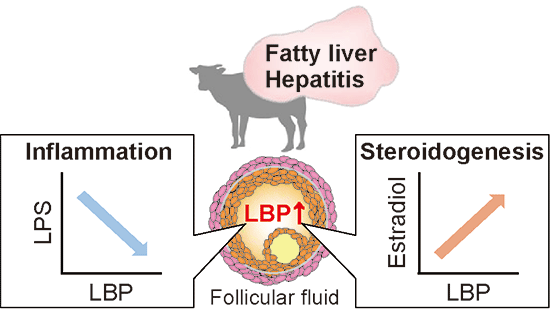 �Graphical Abstract
  �Graphical Abstract
  �Graphical Abstract
  �Graphical Abstract
  扫码关注我们
 求助内容:
求助内容: 应助结果提醒方式:
